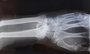
InView Imaging: Your Trusted X-ray Provider
X-rays are essential diagnostic tools used to detect a wide range of conditions, from broken bones to lung infections. At InView Imaging, we prioritize your health and comfort throughout the entire process.
We use state-of-the-art technology to ensure accurate and efficient results. Our experienced radiologists and friendly staff are dedicated to making your experience smooth and stress-free. With convenient locations in Antioch, Oakland, Fremont, and Lafayette, and acceptance of most insurance plans, choosing InView Imaging means choosing exceptional care close to home. Schedule your X-ray today!
Did you know that X-ray technology, radiography, and tomography have been around for over 125 years? This revolutionary imaging technique, such as radiography and tomography, has transformed the fields of medicine, security, and industry with its dimensional image applications. From detecting fractures in bones to screening luggage at airports, radiographs play a crucial role in our daily lives.
X-ray technology allows us to see beyond what meets the eye, providing valuable insights into the human body and various materials. Join us on a journey through the invisible realm revealed by these powerful electromagnetic waves, gamma rays, electrons, and energy.
X-ray Basics
Science Education
X-rays were discovered by Wilhelm Roentgen in 1895, leading to a revolutionary breakthrough in medical imaging. The fundamental principles of x-ray technology involve the interaction of high-energy photons with matter. These photons are generated by accelerating electrons towards a target with a specific atomic number.
The importance of x-rays in modern medicine cannot be overstated. They play a crucial role in diagnosing various conditions, from fractures to tumors, enabling healthcare professionals to make accurate and timely decisions for patient care.
How X-rays Work
X-rays are produced when high-speed electrons collide with a metal target, resulting in the emission of electromagnetic radiation. When these x-rays pass through the body, they interact differently with tissues based on their density and composition. Detectors such as image receptors capture the transmitted x-rays to create detailed images for analysis.
Medical Uses
Diagnostic
X-rays are extensively utilized for diagnosing medical conditions due to their ability to penetrate soft tissues and bones, revealing internal structures. The benefits of using x-rays include quick results and cost-effectiveness compared to other imaging modalities. However, limitations arise when diagnosing conditions that require more detailed views or soft tissue differentiation.
Therapeutic
In radiation therapy, x-rays are precisely targeted at cancerous cells to destroy them while minimizing damage to surrounding healthy tissue. This focused approach helps in eradicating tumors effectively. Despite its efficacy, therapeutic x-ray exposure can lead to side effects such as skin irritation and fatigue.
X-ray Applications
Imaging Bones
X-rays play a crucial role in imaging bone fractures by capturing images of the affected area to determine the extent of the injury. They are essential in assessing bone density by revealing any signs of osteoporosis or bone thinning. The clarity of bone structures in x-ray images allows healthcare professionals to accurately diagnose and treat skeletal issues.
Dental Assessments
In dental examinations, x-rays are utilized to provide detailed images of teeth, roots, and jawbones. Different types of dental x-rays include periapical, bitewing, and panoramic radiographs. These x-rays are vital for identifying cavities, gum disease, and other oral health problems. The importance of dental x-rays lies in their ability to detect oral health issues early, enabling timely treatment and prevention.
Chest Imaging
Chest x-rays are instrumental in diagnosing various lung conditions such as pneumonia, tuberculosis, or lung cancer. They aid in detecting abnormalities like fluid buildup or tumors within the lungs. By providing clear visuals of the chest cavity, these images assist healthcare providers in monitoring respiratory health and guiding treatment plans effectively.
Abdominal Scans
X-rays are commonly used for abdominal imaging to examine organs like the liver, kidneys, and intestines for abnormalities or diseases. Abdominal scans help detect conditions such as kidney stones or gastrointestinal blockages promptly. Patients undergoing abdominal x-ray procedures need to follow specific preparation guidelines like fasting or drinking contrast agents for accurate imaging results.
Understanding Risks
Radiation Exposure
Radiation exposure in x-ray procedures involves the emission of high-energy particles passing through the body to create images. Factors like exposure time and distance affect radiation dose. Minimizing exposure is crucial to reduce health risks.
When undergoing x-ray imaging, the amount of radiation absorbed by the body depends on various factors such as the type of procedure, patient size, and equipment settings. It’s essential for healthcare providers to follow ALARA principles (As Low As Reasonably Achievable) to limit radiation dose while ensuring optimal image quality.
To minimize radiation exposure risks during x-ray procedures, technicians should use proper shielding devices like lead aprons and collars. Patients can also inquire about alternative imaging techniques that involve lower levels of radiation without compromising diagnostic accuracy.
Contrast Medium
Contrast medium plays a vital role in enhancing x-ray images by highlighting specific areas within the body that need closer examination. These agents contain substances that are either radiopaque or radiolucent, aiding in visualizing organs or tissues more clearly.
In x-ray procedures, there are different types of contrast agents used based on the area being examined and the information required from the imaging study. Common types include iodine-based contrast media for vascular studies and barium sulfate for gastrointestinal examinations.
The benefits of using contrast media include improved visualization of structures that would otherwise be challenging to see clearly on standard x-rays. However, potential risks associated with contrast agents include allergic reactions, kidney damage in some cases, and rare instances of severe adverse effects such as anaphylaxis.
Diagnostic Procedures
Identifying Conditions
Kidney Stones
X-rays play a crucial role in detecting kidney stones by capturing images of the urinary tract. These images help healthcare providers identify the presence and location of kidney stones accurately. The effectiveness of x-rays in visualizing kidney stone size and location enables doctors to determine the appropriate treatment plan for patients suffering from this condition.
In diagnosing arthritis, x-rays are essential tools that aid in identifying joint damage caused by the disease. By examining x-ray images, healthcare professionals can observe specific features such as joint space narrowing and bone spurs associated with arthritis. This information is vital for monitoring disease progression over time and assessing the effectiveness of treatment strategies.
Emergency Cases
Swallowed Objects
X-rays are utilized to locate foreign bodies within the digestive system quickly. By performing an x-ray examination, medical professionals can precisely pinpoint the object’s position and determine the best course of action for removal. Safety measures during these procedures include shielding sensitive areas to minimize radiation exposure.
Timely detection through x-ray imaging is critical in emergency cases involving swallowed objects as it helps prevent potential complications such as internal injuries or obstructions. By promptly identifying and locating foreign bodies using x-rays, healthcare providers can initiate appropriate interventions to ensure patient safety.
Therapeutic X-rays
Cancer Treatment
X-rays play a crucial role in cancer treatment by targeting and destroying cancer cells. This therapy is known for its ability to precisely aim at tumors, minimizing harm to surrounding healthy tissues. By emitting high-energy rays, x-ray treatment effectively shrinks or eliminates cancerous growths. The procedure’s success lies in its capability to focus on specific areas affected by cancer.
When treating various types of cancers such as lung, breast, or prostate cancer, x-ray therapy has shown remarkable effectiveness. It involves delivering controlled doses of radiation directly to the tumor site, halting the growth and spread of malignant cells. Patients undergoing this treatment often experience positive outcomes with reduced tumor sizes and improved overall health.
Pain Management
In pain management procedures, x-rays are utilized to provide guidance during interventions aimed at relieving discomfort. These procedures involve pinpointing the exact source of pain within the body using radiography techniques. By visualizing internal structures like bones and joints, healthcare professionals can accurately diagnose conditions causing pain.
The use of x-rays in pain management offers several benefits, including precise localization of issues contributing to discomfort. Through real-time imaging guidance provided by x-rays during treatments such as nerve blocks or joint injections, medical practitioners can ensure accurate delivery of medications for optimal pain relief. This targeted approach enhances patient comfort and improves treatment outcomes.
Technological Advances
NIBIB Research
The National Institute of Biomedical Imaging and Bioengineering (NIBIB) plays a crucial role in advancing x-ray technology. Ongoing research at NIBIB focuses on enhancing x-ray imaging techniques through innovative methods. By collaborating with experts, NIBIB explores new avenues to improve the precision and quality of x-ray imaging.
One significant aspect of NIBIB’s research is its dedication to developing cutting-edge technologies that revolutionize x-ray production. Through meticulous experimentation and analysis, NIBIB scientists are paving the way for more efficient and accurate x-ray procedures. Their work not only benefits medical professionals but also ensures patient safety during diagnostic processes.
Emphasizes innovation in x-ray imaging techniques
Collaborates with industry experts for advancements
Focuses on improving precision and quality of x-ray images
Future Developments
The future of x-ray technology holds promising trends that are set to transform the field significantly. Advancements in x-ray imaging modalities are expected to redefine diagnostic capabilities, offering more detailed insights into various health conditions. These developments aim to make x-rays even more versatile, catering to diverse medical needs effectively.
Innovations in machine learning algorithms may lead to automated interpretation of x-ray results, streamlining the diagnosis process and reducing human error. Enhanced image resolution and 3D reconstruction techniques could provide healthcare professionals with comprehensive views of internal structures, enabling precise treatment planning based on accurate assessments.
Potential for redefining diagnostic capabilities
Automation through machine learning algorithms
Enhanced image resolution for precise treatment planning
Patient Safety
Minimizing Risks
To reduce radiation risks during x-ray procedures, limiting exposure time is crucial. Patients should be positioned accurately to minimize unnecessary radiation. Shielding with lead aprons and collars helps protect sensitive organs.
Optimizing radiation doses plays a significant role in ensuring patient safety during x-ray imaging. Using the appropriate settings on the x-ray machine based on the type of examination minimizes unnecessary exposure. Regular calibration of equipment ensures accurate dosages.
Protective measures such as lead shielding devices and thyroid collars are essential for reducing radiation exposure during x-ray procedures. Proper training for healthcare professionals on positioning techniques also contributes to minimizing risks effectively.
Informed Consent
Informed consent is vital before undergoing x-ray procedures as it empowers patients to make informed decisions about their health. The consent process should include information about the purpose of the procedure, potential risks, and benefits involved.
Patients have the right to understand all aspects of an x-ray procedure before giving consent, including details about potential side effects and alternative options if available. Ensuring that patients are well-informed allows them to actively participate in their healthcare decisions.
Informed consent is not just a formality but a critical step in promoting patient autonomy and safety during medical interventions like x-rays. Patients must receive clear explanations regarding the procedure’s necessity, risks, expected outcomes, and any follow-up care required.
Preparing for X-rays
Before the Procedure
Before undergoing an x-ray, patients need to follow specific preparations to ensure a successful procedure. It is crucial to wear loose, comfortable clothing without any metal objects. Patients must remove jewelry, belts, and other items that could interfere with the imaging process. Moreover, it is essential to inform healthcare providers about any existing medical conditions or pregnancy.
Following pre-procedure instructions is vital for obtaining accurate results during an x-ray examination. Patients may be required to fast for a few hours before the procedure if abdominal imaging is needed. Patients might need to avoid eating or drinking certain substances that can affect the imaging quality.
Informing healthcare providers about relevant medical history such as previous surgeries, implants, allergies, or medications is critical before an x-ray procedure. By providing this information, patients help ensure their safety and prevent any potential complications during the examination.
During the Procedure
During an x-ray procedure, patients can expect a quick and painless process that involves minimal discomfort. Radiologic technologists play a crucial role in conducting x-ray examinations by positioning patients correctly and operating the equipment efficiently. Patients may be asked to hold their breath or stay still momentarily while images are being captured.
To achieve optimal image quality, it is important for patients to follow all instructions provided by radiologic technologists. This includes maintaining proper positioning as directed and cooperating with any breathing instructions given during the procedure.
Aftercare and Results
Understanding Results
X-rays provide detailed images of the body’s internal structures, aiding in diagnosing various medical conditions. Interpreting x-ray images involves identifying abnormalities such as fractures, tumors, or infections. Radiologists play a crucial role in analyzing these images to detect any issues accurately.
Radiologists are specialized physicians trained to interpret medical images like x-rays. Their expertise ensures accurate diagnosis and timely reporting of findings. Once the radiologist analyzes the x-ray, they generate a report detailing their observations for healthcare providers.
Discussing x-ray results with healthcare providers is essential for proper diagnosis and treatment planning. This collaboration ensures that the detected issues are addressed promptly and accurately. Healthcare professionals rely on these reports to determine the next course of action based on the findings.
Follow-up Actions
After receiving x-ray results, it is crucial to follow up with healthcare providers for further guidance. They may recommend additional tests or treatments based on the x-ray findings. Follow-up appointments help in evaluating progress and adjusting treatment plans accordingly.
Follow-up appointments are vital for monitoring recovery progress post-treatment or surgery. Regular imaging through x-rays allows healthcare providers to track changes in conditions over time. This monitoring helps ensure that treatments are effective and adjustments can be made if needed.
X-ray imaging plays a significant role not only in diagnosing but also in monitoring treatment effectiveness over time. Healthcare providers use follow-up x-rays to assess how well treatments are working and make informed decisions about ongoing care.
Summary
You have now gained a comprehensive understanding of x-rays, from their basics to applications, risks, and technological advances. Remember to prioritize patient safety when undergoing diagnostic or therapeutic procedures. Prepare adequately for x-rays, follow aftercare instructions diligently, and discuss your results with healthcare professionals for optimal care. Stay informed about advancements in x-ray technology and how they impact your health.
Keep yourself updated on the latest developments in x-ray procedures and safety measures to make informed decisions about your healthcare. Prioritize your well-being by staying proactive in managing your health and seeking regular check-ups when necessary. Your commitment to understanding and utilizing x-rays responsibly contributes to better healthcare outcomes for you and others.
Frequently Asked Questions
What are the basic principles of X-ray imaging?
X-ray imaging involves using electromagnetic radiation to create images of the inside of the body. It is commonly used for diagnosing fractures, infections, and other conditions by capturing images that show variances in tissue density.
Is it safe to undergo X-ray procedures?
Yes, X-rays are generally safe when performed by trained professionals following strict protocols. The benefits of obtaining crucial diagnostic information often outweigh the minimal risks associated with exposure to ionizing radiation during an X-ray procedure.
How can patients prepare for an X-ray examination?
Patients should wear comfortable clothing without metal accessories and inform the technologist if there is a possibility of pregnancy. They may be required to remove jewelry or metallic objects from the area being imaged for accurate results.
What happens after an X-ray procedure?
After an X-ray exam, patients typically resume their normal activities unless instructed otherwise. Results are interpreted by a radiologist and shared with the referring healthcare provider who will discuss findings and any necessary follow-up steps with the patient.
How have technological advances improved X-ray procedures?
Technological advancements such as digital radiography have enhanced image quality, reduced radiation exposure times, and allowed for more efficient image storage and sharing. These improvements contribute to faster diagnosis, better treatment planning, and enhanced patient care overall.


