General Information and Frequently Asked Questions
If your questions are not answered here, please CONTACT US.
Appointments FAQs
A technologist will come out to escort you to the changing room for your procedure. He or she will be with you throughout your visit. Depending on your procedure, you may be required to change into scrubs. If you are having an MRI, you will remove all metal from your body and store it, along with your personal items, in a secure locker. Next, you will be escorted to the imaging suite where the technologist will explain the details of your exam and give you an opportunity to ask questions before performing the procedure.
We perform x-rays during all business hours on a walk in basis. However, if you have a referral for several x-rays, you may schedule an appointment. Mammograms, ultrasounds, CT, MRI, DEXA (bone density) and interventional procedures do need an appointment as they often require preparation.
Most, but not all, imaging studies require a physician referral. Body composition exams do not require a referral. Please inquire about the specific examination if needed.
Yes. You may choose any facility to have the examination performed.
Bone Denisty DEXA Scans FAQs
Osteoporosis occurs when bones lose their density, become weak and fragile and thus more likely to fracture. Bone density reaches a peak in your 20’s, and declines at a variable rate thereafter. Osteoporosis commonly affects women, particularly after menopause, although men and teenagers may also be affected. Activity and a healthy diet rich in calcium and vitamin D has been shown to help prevent osteoporosis.
Osteoporosis is often called “silent” as there is no pain until the bone actually breaks. The hip, spine and wrist are most susceptible to fractures. Many physicians will recommend a DEXA exam for women approaching menopause to evaluate bone strength, and then periodically afterward to monitor your bone health. Your physician may elect to treat you if your bone density is very low.
CT Scans FAQs
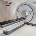
CT (computed tomography), sometimes called CAT scan, uses special x-ray equipment to obtain image data from multiple different angles all around the body and then uses computer processing of the information to show a cross-section of body tissues and organs.
CT imaging is particularly useful because it can show several types of tissue- lung , bone, soft tissue and blood vessels-with great clarity. Using specialized equipment and expertise to create and interpret CT scans of the body, radiologists can more easily diagnose problems such as cancers, cardiovascular disease, infectious disease, trauma and musculoskeletal disorders.
Because it provides detailed, cross-sectional views of all types of tissue, CT is one of the best tools for studying the chest and abdomen. It is often the preferred method for diagnosing many different cancers, including lung , liver and pancreatic cancer, since the image allows a physician to confirm the presence of a tumor and measure its size, precise location and the extent of the tumor’s involvement with other nearby tissue. CT examinations are often used to plan and properly administer radiation treatments for tumors, to guide biopsies and other minimally invasive procedures and to plan surgery and determine surgical resectability. CT can clearly show even very small bones as well as surrounding tissues such as muscle and blood vessels. This makes it invaluable in diagnosing and treating spinal problems and injuries to the hands, feet and other skeletal structures. CT images can also be used to measure bone mineral density for the detection of osteoporosis . In cases of trauma CT can quickly identify injuries to the liver, spleen , kidneys or other internal organs. Many dedicated shock-trauma centers have a CT scanner in the emergency room. CT can also play a significant role in the detection, diagnosis and treatment of vascular diseases that can lead to stroke , kidney failure or even death.
You should wear comfortable, loose-fitting clothing for your CT exam. Metal objects can affect the image, so avoid clothing with zippers and snaps. You may also be asked to remove hairpins, jewelry, eyeglasses, hearing aids and any removable dental work, depending on the part of the body that is being scanned. You may be asked not to eat or drink anything for one or more hours before the exam. Women should always inform their doctor or x-ray technologist if there is any possibility that they are pregnant.
Ultrasound FAQs
Ultrasound can be used to safely exam babies before they are born. Early in pregnancy ultrasound is used to confirm the well being of the fetus before you can feel the baby moving and before your doctor can hear the heartbeat. After about 16 weeks, as the internal organs and bones are developing, ultrasound can assess the fetal growth and development. Examining babies while in the womb is completely safe and the information provided can be very helpful to your doctor.
Ultrasound may be used to evaluate any of the internal organs in your body, and your exam will be tailored to focus on the organs of interest as requested by your physician. We may focus on the gallbladder if you have right upper quadrant pain, the liver if you have abnormal blood tests or a history of hepatitis, or the kidneys if you have blood in your urine or pain in your side. In the pelvis, we may examine the uterus, endometrium or ovaries if you have pelvic pain, cramping or abnormal bleeding.
Doppler ultrasound examines moving blood within your arteries and veins and creates real-time images which can be used to search for narrowed or blocked vessels, aneurysms and blood clots. Doppler ultrasound is most commonly used to evaluate the arteries leading to your brain or search for abnormal enlargement (aneurysm) of your aorta.

Health Screenings
Screening involves searching for disease before symptoms are present and is usually performed because you have increased risk of developing a disease. Modern imaging equipment is very sensitive at detecting certain diseases such as arterial blockage, aneurysms, cirrhosis and cancers before you have symptoms.
It is important to remember that your doctor is not requesting the screening test because he/she has clues that you have a disease. Rather, they may recommend a test due to your family genetic background, risk factors such as smoking or poor diet, or findings on your blood tests. It is also important to know that these tests often detect other insignificant findings which will not impact your health but will be noted in your report.
We require that you have a physician to receive the screening exam results. These test results are most useful in the hands of well trained physicians who understand the significance of the findings. Should your test reveal an abnormality that might impact your health, your physician may choose to order additional tests. These tests do not take the place of routine physician health check-ups.
We offer many different kinds of Health Screens and Health Screening Packages. See our list below.
Health Screening Services:
Carotid Artery Screen (US)
Aortic Aneurysm Screen (US)
Lung Cancer Screen (CT)
Virtual Colonoscopy Diagnostic or Screening (CT)
Whole Body Scan (MRI/CT)
Abdomen, Pelvis, and Brain MRI Package
Abdomen, Pelvis, and Brain MRI with Chest CT Package
Abdomen, Pelvis, Brain, and Chest CT Package
Recent advances in Magnetic Resonance Imaging (MRI) have allowed for fast, reliable and safe disease screening for detecting diseases throughout the body. MRI is far safer than Computed Tomography (CT) scanning. The magnetic field and radiofrequency energy of MRI is safe. In recent years, disease screening using CT technology has been prevalent, but there are many negative considerations when using CT to scan the entire body. One of the most important reasons is due to the amount of radiation exposure.
With your safety in mind, we have created a disease screening method using a combination of MRI and CT, in order to limit radiation exposure as much as possible. Most other centers advertise body scans using only CT technology because it is much faster but the amount of radiation exposure from scanning an entire body may be harmful.
People with a family history of cancers such as lung, lymph, stomach, kidney, ovarian and liver cancer or who have had exposure to environmental toxins may be good candidates for this type of screening exam.
A painless test recommended by the American Heart Association as part of routine risk assessment for detection of heart disease now also is recognized as an important tool to detect stroke risk.
Carotid Screening is a good risk assessment for the detection of heart disease. This painless test uses ultrasound to detect fatty plaque buildup in the carotid arteries on each side of the neck. Plaque in these arteries is a good indicator of arterial health throughout the body and an even more direct direct evaluation of the risk of stroke.
An abdominal aortic aneurysm (AAA) is an enlargement and weakening of the aorta which can leak or rupture and cause life-threatening bleeding. It is usually asymptomatic and is often found incidentally during testing for other conditions. When detected early, surgery can be performed to repair an AAA defect before it becomes a problem. Some Medicare recipients may be eligible for this screen to be covered under their insurance.
Lung cancer is the number one cause of cancer death in American men and women, killing approximately 160,000 each year. If the diagnosis is made early (stage I disease), the percentage of those alive five years after diagnosis is 60-80%. Unfortunately, lung cancers are often not detected until after symptoms appear, when the lung cancer is already at later stages and the cure rates are significantly lower. The percentage alive in five years after a diagnosis of advanced disease is only 1-13%. Without lung cancer screening, the percentage of lung cancers diagnosed with stage I disease is only 15%. In a population of people screened yearly for lung cancer with CT, the percentage of cancers diagnosed with stage I disease rises to 75%.
Five-year survival rates are clearly different for diagnoses made at different stages. However, there is some ongoing debate in the medical community and ongoing research as to whether screening for lung cancer is cost effective and whether detecting lung cancer at an earlier stage really makes a difference in overall survival. It is possible that people diagnosed with lung cancer at an early stage using CT screening will still die at the same time they otherwise would have, except with a longer lead time between when they know of their disease and when they die. Some argue that survival rate at five years after diagnosis is higher because of “lead time bias.” This argument is that the numbers only seem better because we start counting the five years at an earlier time, and not because earlier diagnosis lead to more effective intervention. The fact that the number of lung cancers diagnosed each year is almost identical to the number of deaths from lung cancer each year (within 10%), suggests that almost everyone who gets lung cancer, even those diagnosed with early stage disease, dies of it. While it seems intuitively obvious that earlier diagnosis should allow higher cure rates, the debate will continue for some time, until more definite long-term follow up research data is available.
According to the American Cancer Society, colorectal cancers are the second most common cancers in men and women. Over 60,000 Americans die from colon cancer every year, yet colon cancer is one of the most treatable forms of cancer if caught early on.
If you are one of those that avoid this important screening because you are embarrassed or afraid, Virtual Colonography (VC) may be just for you. VC is also useful when conventional colonoscopies cannot be completed due to strictures or other forms of pathology.
Virtual Colonoscopy allows patients a less-invasive and far more comfortable exam without the need for sedation, and a minimal interruption to the day. It is also safer than traditional methods because it is less likely to cause perforations or damage to the colon because it does not require the use of a colonoscope. No recovery period is required since sedation is not used during this exam and patients are able to return to normal activities immediately afterwards. Actual scan time is about 10 minutes. After the scan, 3D images will be created, which our radiologist will review for abnormalities.
The American Cancer Society recommends that screening for colorectal cancer begin at age 40 or older. People should begin colorectal cancer screening earlier and/or undergo screening more often if they have a family or personal history of colorectal cancer, adenomatous polyps or chronic inflammatory bowel disease.
Imaging Results
Our technologists are experts at obtaining diagnostic images and will let you know that an exam is complete. However, technologists or other staff members are not trained to read radiology exams and therefore cannot provide any results.
We strive to interpret your exam and get results to your referring physician as fast as possible, 2-3 business days. If marked urgent, we will call or fax your results promptly. Your doctor will then contact you regarding the results. If interpretation requires prior imaging studies or additional clinical information, your results may take longer as we wait for prior imaging studies.
Once your exam has been interpreted and sent to your referring physician, you may request a copy of your report for personal use. You will need to sign a medical release to receive the report. All mammography patients receive a results letter in easy-to-understand language, either upon completion of your exam or within 2 weeks.
All physicians have direct portals to our images, which they may view within minutes of completion. For a small fee and a signed medical release, you may obtain a CD with your images. If you have any questions, please contact our Medical Records Department at the appropriate office.
We retain our records beyond the state and federal guidelines, which are 7 years or until minor patients turn 18 years old.

Insurance
There are thousands of individualized insurance plans. Many plans have rigid medical necessity requirements for certain exams, and some plans have strict time guidelines for screening exams such as mammography. We will do our best to answer your insurance questions, but the responsibility is ultimately yours and we recommend that you check with your insurance plan to assure coverage. Please call the 800 number located on the back of your insurance card and speak with member services to assist you with individual coverage information.
Most insurance companies require pre-authorization for more complicated exams, particularly MRI and CT scans. Although it is the responsibility of your referring doctor to obtain the necessary authorization, we provide this service for certain exams free of charge to your physician. If the exam is performed without prior authorization, we cannot assure payment from your insurance company.
If you have a doctor’s referral slip, you may have a medical diagnostic exam performed. We do offer a discount for exams paid at the time of service, but please inquire at the front desk prior to service.
Yes! You are not obligated to go to a specific facility. Even though your doctor may recommend one, ultimately, you should feel comfortable with the facility and have trust in the medical personnel providing service. Since a radiologist is a highly trained and specialized physician, you should choose your radiologist just as you would carefully select a surgeon or internist.
Feel free to inquire about the cost of the test as it can vary significantly. For example, hospitals and their outpatient sites routinely charge several times as much for the same exam or procedure performed at an independent outpatient imaging center. You may be quite surprised at the difference in cost. And remember that even if you have health insurance, your may incur fees through co-pays or unmet deductibles.
Quality
Yes, we maintain accreditation by the American College of Radiology (ACR), the most stringent credentialing body, who periodically evaluate equipment, personnel and quality assurance procedures. This rigorous process assures that we have met nationally accepted standards regarding MRI, CT, ultrasound and mammography. We also satisfy obligatory State and Federal x-ray, mammography and other imaging guidelines to assure safety and quality control.
Radiation
We only perform x-rays when medically necessary, use as little radiation as possible to evaluate the area of concern and shield other areas when possible. Your physician believes this exam is important for your diagnosis and treatment.


