Key Takeaways
-
The FDA’s recent updates aim to modernize mammography by introducing new assessment categories, mandatory tissue density reporting, and enhanced quality standards. These changes are designed to improve early breast cancer detection and streamline patient care.
-
Additional imaging technologies, such as 3D mammography (tomosynthesis), molecular breast imaging (MBI), and contrast-enhanced mammography (CEM), improve detection modalities. These failures lead to less-accurate outcomes for breast screening. They lower false positives, enhance clarity, and help spot cancers that 2D mammograms miss.
-
Artificial intelligence (AI) has begun to revolutionize mammography by helping physicians analyze images, enhance diagnostic accuracy, and standardize image evaluations. AI is another key ingredient to helping optimize workflows and AI will be a great tool to help radiologists find abnormalities with more accuracy than ever before.
-
ABUS use, especially alongside 3D mammography, provides an important new resource for women with dense breast tissue. It works alongside standard mammography to deliver better overall screening and improve the detection of difficult-to-find cancers.
-
Recent mammography advancements focus on patient safety and comfort. They work to limit fear, eliminate pain, and inform patients about the mammography experience. These advancements promote increased screening compliance and more positive experiences overall.
-
Inclusive access to this new mammography standard is still very much a priority, with mobile units, telehealth, and community outreach programs aiming to connect with underserved populations. Advocacy for insurance coverage and cost transparency is critical to ensure equitable access to these, and other emerging life-saving technologies.
One of the biggest recent advancements in mammography is the introduction of this 3D imaging technology, referred to by the name digital breast tomosynthesis (DBT). This new technology produces an extensive series of images, so physicians can review breast tissue one layer at a time.
Unlike traditional 2D mammograms, 3D mammography allows your doctor to reduce overlapping tissues, improving accuracy in detecting abnormalities and reducing the need for callbacks. It’s especially advantageous to women with dense breast tissue, providing them with greater clarity, fewer false positives, and faster detection of problems when found on a mammogram.
Recent advances in artificial intelligence (AI) have revolutionized the image analysis playing field. This innovation is now enabling radiologists to more quickly and accurately detect subtle signs of breast cancer.
By making the screening process more accurate, these innovations result in better outcomes for patients. They do this all while emphasizing comfort and compassion every step of the way.
What is New in Mammography?
The field of mammography continues to evolve with advanced technologies and updated regulatory standards aimed at improving breast cancer detection and patient outcomes. The FDA recently introduced significant updates to modernize breast cancer screening, the first in over 20 years.
These include mandatory reporting of breast tissue density, which informs patients of factors that may obscure cancer detection, and the addition of new assessment categories to refine diagnostic precision. Modernized quality standards are expected to improve the consistency and reliability of mammography practices, ensuring earlier detection and more personalized care.
1. Understand 3D Mammography (Tomosynthesis)
3D mammography, or digital breast tomosynthesis (DBT), provides a more comprehensive look at breast tissue. It takes several images from different angles to form a three-dimensional view.
This method minimizes visibility problems with dense, overlapping tissue that are typical with 2D imaging, improving cancer detection rates. Research studies indicate that the Selenia Dimensions from Hologic detects between 20-65% more invasive cancers.
This is particularly the case for women who have dense breasts. By providing clearer imaging, DBT reduces the need for unnecessary biopsies, making it the best choice for precise screening.
2. Molecular Breast Imaging (MBI) Details
MBI utilizes a specialized radiotracer to illuminate cancerous cells, providing information in areas that traditional mammograms are unable to detect. This approach works best in dense breast tissue, where older and more traditional approaches have a harder time detecting cancer.
Enabling personalized screening strategies by identifying cancers at significantly earlier stages, it adds a new imaging modality to current clinical applications.
3. Contrast-Enhanced Mammography (CEM) Overview
Contrast-enhanced mammography (CEM), which includes injecting a dye, increases imaging contrast and focuses on suspicious lesions. Cost-effective solution CEM is the safest solution as it could avoid unnecessary additional testing to detect abnormalities.
It is a useful substitute for MRI, particularly in high-risk patients.
4. AI in Mammography: Current Applications
AI supports mammography through accurate classification of breast density, improving image analysis and decreasing radiologist burnout. It remains promising to further standardize such evaluations, integrating findings across imaging modalities and thereby improving the strength of evidence.
5. Update on Automated Breast Ultrasound (ABUS)
ABUS allows for a gentler approach to 3D imaging, particularly advantageous for women with dense breast tissue. When used in conjunction with traditional mammography, it outperforms them in detecting cancers at early, more treatable stages and overall screening results.
How Does 3D Mammography Work?
3D mammography, or digital breast tomosynthesis (DBT), is a revolutionary new breast cancer screening tool. This state-of-the-art technology enhances conventional mammograms by capturing several pictures of breast tissue from different angles.
It then uses those images to mathematically reconstruct into a clear, three-dimensional view. 3D mammography creates sharper, more detailed images. In doing so, it’s able to catch breast cancer sooner, sometimes as much as two years before a tumor can be felt, and finds smaller abnormalities that didn’t appear on traditional mammograms.
During this process, rates of invasive breast cancer detection jump by as much as 40%. It reduces false positives, so you can worry less.
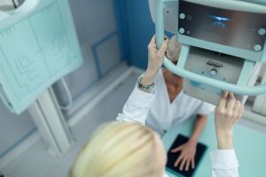
Image Acquisition Process Explained
First, we take the patient’s breast and place it on an angled platform. Next, we use mild compression to keep tissue evenly distributed so we can get the clearest images possible.
The 3D mammography machine moves like a pendulum over the breast, taking hundreds of low-dose X-ray images from different angles. This entire procedure only takes 10 to 15 minutes – similar to a conventional mammogram.
The same equipment utilizes state of the art detectors to deliver exceptional image quality with the lowest possible radiation dose. Appropriate patient positioning and technique are imperative. This is important because it helps us get complete visualization of the breast tissue, including denser areas where abnormalities may be hidden.
Reconstructing 3D Images from 2D
The resultant slices are then reconstructed by complex algorithms that layer the 2D slices together into a high-resolution 3D image. This process allows radiologists to view breast tissue one layer at a time.
Consequently, it greatly decreases the masking effect of superimposed tissue. This clarity immeasurably improves diagnostic precision, allowing for the identification of the smallest and most malignancies.
Reading and Interpreting 3D Mammograms
Expert radiologists interpret these reconstructed images on a dedicated reader with proprietary processing techniques. Because 3D mammograms are more detailed, they need to be read by someone specifically trained, unlike when reading 2D results.
This detailed, 3D view results in fewer false positive and negative diagnoses, aiding in early treatment decisions when breast cancer is most successfully treated.
Benefits and Drawbacks of 3D Imaging
3D mammography, known as digital breast tomosythesis, is the biggest breakthrough in breast cancer detection in decades. It provides an unbiased, thorough, and multi-layered view of breast tissue, allowing for higher quality diagnoses. This cutting-edge solution promises to solve many well-known challenges in conventional imaging, providing increased clinical confidence as well as a better experience for patients.
Yet, it equally brings forth issues that should be considered to better appreciate its overall effect.
Improved Detection Rates Discussed
In large trials, 3D mammography has made incredible strides in detecting cancers early. Research shows as much as a 41% increase in detecting invasive breast cancers over 2D imaging. When it comes to fighting cancer, catching it at the very beginning is essential.
It sometimes results in more conciliatory courses of action, such as forgoing chemotherapy altogether, and achieves higher rates of survival. Women of all ages receive tremendous benefits from this precision. This can particularly be the case for patients with dense breast tissue, which can obscure potential abnormalities on traditional imaging.
Young women and patients with a heritable family history of breast cancer will see the most benefit, Margulies said. They, in turn, can expect more customized treatment approaches.
Reduced False Positives Explained
The biggest benefit is the dramatic decrease in false positives. This advancement reduces the emotional burden and need for additional follow-up procedures. In fact, one study showed that there was a 3.4 percent decrease in callbacks when adopting 3D imaging.
This decrease results in less redundant testing, such as ultrasounds or MRIs, which can be expensive and time-consuming. This precision lowers healthcare costs and lessens patient stress, improving the quality of care received.
Radiation Exposure Considerations
3D mammography addresses worries about radiation exposure. It operates at doses that are similar, and in some cases lower than 2D mammography. Safety protocols keep exposure well below hazardous levels, making this technology powerful and safe.
Cost-Effectiveness Analysis
While 3D mammography is more expensive upfront, there is a clear benefit. First, by detecting cancer earlier, it reduces long-term healthcare costs by lowering the need for invasive treatments and additional imaging tests.
For patients and health care systems alike, this means saving money. Equally important is insurance coverage for 3D imaging — without it being covered, it cannot be made widely available.
Comparing 2D and 3D Mammography
This technological shift from traditional 2D mammography to advanced 3D mammography is known as digital breast tomosynthesis and greatly improves breast cancer detection. These benefits to patient care are at least as important as the innovative edge of this advancement. Knowing how these hands work differently brings more attention to what each one does best for breast health.
|
Feature |
2D Mammography |
3D Mammography |
|---|---|---|
|
Detection Rate |
Standard detection capability |
Detects up to 40% more cancers |
|
Imaging Technique |
Flat, two-dimensional images |
Layered, three-dimensional images |
|
False Positives |
Higher rate |
Reduced rate |
|
Screening Time |
~33 seconds |
~77 seconds |
|
Cost |
Typically lower |
Generally higher |
Advantages of Traditional 2D Mammography
Regular traditional 2D mammography is still a fundamental, proven and trusted screening tool. Its legacy as the first line of defense against breast cancer paved the way for more advanced and innovating imaging technologies today.
Its familiarity makes it highly accessible, ensuring availability in almost any healthcare setting, and providing a consistent patient experience.
2D mammograms are typically less expensive, which can make them a cost-effective option for regular, preventive screenings.
Limitations of Traditional 2D Mammography
While 2D mammography has its advantages, it does have its shortcomings. Because dense breast tissue is particularly effective at hiding cancerous lesions, this results in missed diagnoses.
It comes with a significantly higher rate of false positives, often leading to unnecessary follow-ups. Additionally, radiologists might have a difficult time interpreting overlapping breast structures, further heightening the need for supplemental imaging.
When is 2D Mammography Still Appropriate?
2D mammography continues to be a cost-effective and scientifically valid standard of care for routine screening in average-risk patients.
It performs suitably enough in environments lacking access to high-quality technology or where high expense is prohibitive. For others, it simply enhances 3D imaging to provide a better, more complete evaluation.
Early Detection: Why It Matters
Early detection is key to saving lives through the successful treatment of breast cancer. Finding cancer when it’s most treatable means patients can receive effective treatment as soon as possible. This proactive approach allows for better patient outcomes and reduces the physical burdens associated with more advanced therapies.
Decades of research unequivocally demonstrate that early detection saves lives by dramatically lowering mortality rates as you will see below. Since the advent of the randomized control trials in the 1960s, these trials have proven a 20-40% reduction in deaths due to mammographic screening.
Even with amazing new treatments, more than 40,000 women in the U.S. Die from breast cancer each year. This shocking statistic means we need to take more proactive action now.
Impact on Treatment Outcomes
When detected early, breast cancer often requires less aggressive treatment options, such as localized surgery or targeted therapies, minimizing side effects and recovery times. Research has just about across the board proven that patients whose cancers were detected earlier are more likely to survive and lead long-term healthy lives.
Beyond clinical benefits, early detection provides psychological reassurance, offering patients a greater sense of control and reducing anxiety associated with advanced treatment stages. Early detection is especially important to all people with dense breast tissue.
In such scenarios, mammographic sensitivity may be reduced, resulting in missed diagnoses.
Screening Guidelines and Recommendations
National guidelines call for mammographic screenings starting at age 40 for those of average risk, with tailored screening schedules for those at increased risk. The American Cancer Society and other leading organizations recommend personalizing timelines.
They calculate these changes based on the trajectory of medical progress, independent risk factors, and every patient’s pertinent history. Consistently revising guidelines keeps them in line with advancements, like 3D mammography, that help correct shortcomings in older practices.
Risk Factors and Personalized Screening
Personalized screening strategies take into account genetic predispositions, lifestyle factors, and the presence of dense breast tissue. Reassuringly, genetic testing—especially for BRCA1 and BRCA2 mutations—was able to identify these high-risk individuals, thus enabling them to have screenings earlier and more regularly.
Lifestyle considerations, such as diet and physical activity, influence screening decisions too, highlighting the benefits of high-touch, personalized care.
Accessing Advanced Mammography Options
Advanced mammography technologies are helping to revolutionize how we find breast cancer. Their adoption has been increasing rapidly across all types of healthcare environments. Digital breast tomosynthesis (DBT) has quickly become the standard of care in the U.S. Healthcare professionals overwhelmingly acknowledge its unmatched diagnostic performance.
Facilities providing 3D mammography represent more than 60% of the facilities, a result of the rapid adoption of this technology. DBT has received FDA approval for use in women with dense breast tissue. It is a much better alternative than 2D imaging alone, finding 20 to 65% more invasive cancers.
Availability in Different Healthcare Settings
Advanced mammography services are available in hospitals, specialty imaging centers, and private practice clinics. Yet, huge gaps remain in access between cities and rural communities. Urban centers usually have a much larger concentration of facilities outfitted with the most advanced systems such as Hologic’s 3Dimensions.
Just as important, rural areas are seeing the benefits of successful collaborations between hospitals and imaging centers. Telehealth consultations are fueling this success, providing specialized expertise at a distance. These partnerships help provide greater geographic reach and increase access to the most advanced screenings available.
Insurance Coverage and Costs
Insurance is the key player in providing access to advanced mammography. Even though all providers must cover routine mammograms, coverage for 3D imaging can vary widely. Patients should be proactive and review their insurance plan details to get a better idea of what they may need to pay.
Ongoing advocacy efforts continue to call for more expansive coverage. We want to lower the financial burdens to patients and make sure everyone has equitable access to these advanced options.
Overcoming Barriers to Access
Mobile mammography units have proven to be indispensable resources in accessing populations in need, especially individuals in rural or lower economic communities. By taking screening into the heart of communities, these units remove other logistical barriers including transportation.
Community outreach programs create spaces for awareness, and patient education initiatives help women feel confident in making regular screenings a priority.
The Role of AI in Mammography
Artificial intelligence (AI) is transforming mammography by increasing diagnostic accuracy, improving workflows, and redefining breast cancer screening. Infusing this kind of technology into daily imaging practice changes the game for radiologists and patients alike. It addresses some of our most stubborn issues with accuracy and efficiency.
AI-Assisted Image Analysis
AI systems aid radiologists by analyzing mammographic images at an extraordinary speed and accuracy. These tools are best at detecting subtle abnormalities. They are able to find microcalcifications and small masses that are lost in the noise of human analysis.
For instance, AI implementation improved the detection of small cancers from 36.6% to 44.9%, showcasing its ability to identify earlier-stage cancers. In its third study, AI decreased the false-positive rate by 32%, reducing the risk of giving unnecessary stress to patients and lowering recall rates by 20.5%.
By automating repetitive tasks, AI reduces the radiologists’ workload by 33.5%, giving them more time to focus on complex cases. This partnership-focused method maximizes resources and enhances patient care and outcomes dramatically.
Improving Accuracy and Efficiency
AI augmentation improves the accuracy of mammogram readings with higher precision, boosting the detection rate of invasive cancers from 79.6% to 84.9%. Its advanced algorithms can detect even the slightest changes in breast tissue, giving you a head start on early diagnosis.
Efficiency gains are just as impressive, with AI streamlining image processing, processing and interpretation, expediting time to care. For women who have fatty breasts, AI has increased cancer detection rates from 0.52% to 0.68%, making this technology all the more dependable.
Following up on patient care is improved by the increased accuracy, which allows for more direct interventions and modified care recommendations.
Future Potential of AI in Screening
All in all, the future of AI in mammography looks bright. Ongoing research aims to refine algorithms, enabling personalized screening strategies based on individual risk profiles.
These innovations have the potential to remake our healthcare systems into more cost-effective enterprises that increase access to long-term, high-quality screening.
Patient Safety and Comfort Improvements
Enhancements in mammography improve patient safety and comfort, making significant strides in resolving common concerns that have prevented people from seeking regular screenings. These safety and comfort improvements further enhance the pedestrian experience. They enhance patient emotional comfort, inviting greater numbers of them to engage in this extremely important diagnostic experience.
Reducing Anxiety During Screening
Decreasing patient anxiety is key to creating a positive screening experience. Straightforward communication from healthcare professionals involved in an inpatient or outpatient procedure is crucial, helping patients know what to expect at every stage of the process. Factors that create supportive environments, including warm lighting and private spaces, are central to fostering a calming atmosphere.
Relaxation methods, such as guided breathing exercises, can help you relax even more. Patients with claustrophobia frequently elect for contrast-enhanced mammography (CEM) rather than conventional breast MRI. They love the faster processes, quieter operations, and less stressful environment that CEM has provided.
CEM, with its rapid acquisition times, upholds diagnostic interpretive quality making it a strong alternative for patients who have difficulty with MRI.
Minimizing Discomfort Techniques
Technological innovations have done a lot to lessen pain associated with mammograms. Today’s state-of-the-art imaging equipment, 3D mammography included, uses less compression. It incorporates more advanced positioning techniques to allow for a more comfortable experience for patients.
In addition to physical strain, proper alignment by highly-trained technicians greatly reduces risk. Digital mammography allows for more precise imaging and less discomfort for patients. AI technology enables radiologists to detect abnormalities with 41% increased accuracy compared to traditional 2D techniques.
More efficient procedures, such as CEM, improve patient comfort by asking for fewer seconds without sacrificing quality.
Addressing Patient Concerns
Patients have a lot on their minds heading into a complicated procedure—including pain, precision, and risk all among them. Teaching them more about advanced technologies such as CEM, which utilizes CT-based contrast agents to produce detailed imaging, is a way to calm apprehensions.
Finally, when providers approach misconceptions with thoughtful, individualized guidance and the establishment of feedback loops, it builds trust. In addition, digital imaging’s ability to quickly share results for second opinions further reassures patients, having a positive impact on overall patient satisfaction.
Future Trends in Mammography
Future improvements in mammography are sure to continue expanding the paradigm of breast cancer detection, improving detection accuracy and patient care through greater integration with personalized medicine. Advancements like these work to overcome shortcomings of conventional screening methods while improving early detection and treatment prognosis.
Emerging Technologies on the Horizon
Digital mammography and 3D tomosynthesis represent only the beginning of a full-scale revolution as discovery of new technologies continues to transform mammography. The additional images created by 3D tomosynthesis, for example, allow the technique to be very effective in catching cancers that are otherwise obscured by dense breast tissue.
Contrast-enhanced mammography (CEM) is a major innovation in breast imaging. By injecting iodine contrast to highlight abnormalities, it increases sensitivity from 80% to almost 100%. Artificial intelligence (AI) and machine learning are increasingly utilized to detect patterns in mammographic images, aiding in predicting cancer risk with precision.
Research into these technologies is ongoing, with institutions exploring innovative imaging techniques to reduce false negatives and improve diagnostic consistency. By adding more accuracy and reliability to the screening process, these advancements promise to help patients receive better care.
Personalized Screening Approaches
Screening targeted to specific individual risk factors is underway and increasing. By integrating genetic testing, family history, and lifestyle factors, we can provide personalized recommendations that lead to higher early detection rates.
This transition is particularly significant for patients with dense breast tissue. The FDA requires that all of these patients be informed about the potential negative impact of this on imaging quality. Finally, personalized approaches help to engage patients in their own care, making people more informed about their individual risk profile and screening requirements.
Integration with Other Diagnostic Tools
Additionally, integrating mammography with other imaging modalities, including MRI or ultrasound, provides a fuller diagnostic procedure. Multidisciplinary approaches improve accuracy, particularly for difficult cases.
This multidisciplinary integration leads to synergistically improved outcomes by using each modality’s complementary strengths, increasingly resulting in improved patient care and treatment planning.
Conclusion
Innovations in mammography promise to deliver tangible gains in early detection and improved patient experience. Because 3D imaging allows doctors to see more detail, they are better able to identify smaller and more difficult-to-detect abnormalities. AI tools provide another layer of accuracy, increasing analysis speed while lowering the potential for human error. These enhancements result in less missed cancer and false positives, meaning screenings are more effective and reliable.
Beyond technological advancement, comfort and safety have grown by leaps and bounds for patients. Thanks to improved equipment design and more personalized care, the experience is less intimidating. Even as access to these tools expands, awareness and advocacy are still crucial to breaking down barriers.
Understanding why these new advancements are important is key. We know regular screenings can save lives, but with these technologies available, they can save even more. Discuss these new advancements with your doctor to find the right option for you. When it comes to your health, seize the day—early detection is key.
Frequently Asked Questions
What is the newest advancement in mammography?
The newest advancement, 3D mammography or digital breast tomosynthesis, helps to overcome these challenges. It produces high-resolution, 3-dimensional, layered images of the breast distributing tissue and abnormality, increasing accuracy in finding abnormalities.
How does 3D mammography improve early detection?
It offers a better, deeper look into breast tissue. This improves outcomes by catching smaller cancers at an earlier stage, particularly in dense breast tissue.
What are the benefits of 3D mammography over 2D?
One of the key benefits of 3D mammography is a decrease in false positives and unnecessary callbacks. It delivers a lower radiation dose while increasing cancer detection rates by capturing multiple angles to produce more precise imaging.
Is 3D mammography safe?
Answer: Yes, mammography is safe. It uses low-dose radiation similar to 2D mammography but with better visualization.
How does AI enhance mammography?
AI is another tool to assist radiologists, analyzing images at a speed and detail that can quickly highlight potential abnormalities. This minimizes human mistakes and increases the overall diagnostic precision rate.
What improvements have been made for patient comfort?
With improved mammography machines using softer paddles and faster imaging, along with some procedural improvements, discomfort experienced during the test can be minimized. These days, many clinics are working—informed by the rise of the “patient experience”—to present a more inviting, patient-friendly front.
What are future trends in mammography?
Future advancements include AI integration, better imaging for dense breast tissue, and portable mammography devices to improve access for underserved populations.