Key Takeaways
-
Advanced high-field MRI is state-of-the-art imaging technology. It uses high-field magnets to provide comprehensive imaging that exceeds conventional techniques and improves diagnostic confidence.
-
High-field MRI refers to magnetic field strengths higher than 1.5T. This technology offers better image quality, increased detection of abnormalities, and increased visualization of dense breast tissue when compared to conventional MRI.
-
Breast MRI plays an increasingly important role in early cancer detection, especially for women at high risk. It performs an irreplaceable role in evaluating tumors, directing treatment, and tracking changes in breast tissue.
-
Using mammography in conjunction with breast MRI increases the rate of detecting cancers. This is particularly the case when mammograms are inconclusive, allowing for a more complete and efficient diagnostic process.
-
Preparing for a breast MRI involves following pre-scan instructions, such as avoiding specific medications or foods, arriving early, and communicating any concerns with medical staff to ensure a smooth procedure.
-
Today’s MRI machines enhance patient comfort with roomier designs and noise-reduction technology. Reducing scan times makes the procedure less daunting and anxiety-producing.
Advanced high-field MRI for mammograms offers a detailed and precise imaging solution for breast cancer detection and diagnosis. Using magnetic fields stronger than standard MRI systems, high-field MRI captures sharper and more accurate images of breast tissue.
This method provides enhanced visualization of abnormalities, making it a valuable tool for detecting early-stage cancers and assessing dense breast tissue. It reduces the need for invasive procedures by offering clear imaging, supporting better clinical decisions.
High-field MRI is particularly beneficial for patients with a higher risk of breast cancer or complex cases requiring detailed evaluation. By integrating this advanced technology, healthcare providers can improve diagnostic accuracy and offer more personalized care for patients.
What Is Advanced High-Field MRI
Advanced high-field magnetic resonance imaging (MRI) represents one of the most substantial advances in medical imaging. It employs high magnetic fields to produce incredibly detailed and accurate images. This technology features magnetic field strengths greater than 1.5 Tesla (T). Consequently, it offers the highest clarity and disease-detecting ability available through any other imaging modality.
It is most capable of portraying complex details, which makes it a fundamental tool in diagnostic medicine. It aids the detection of subtle abnormalities and contributes to higher diagnostic accuracy, even in more complex conditions.
Definition of High-Field MRI
High-field MRI are imaging systems with magnetic field strengths starting at 1.5T. These systems are capable of ultra-high fields of 7T or 8T. These systems are very different from conventional MRIs, which are often at lower field strengths like 0.5T or 1.0T.
The higher field strength has a direct impact on the signal-to-noise ratio (SNR), resulting in clearer, crisper and finer detailed images. At 8T, and a matrix size of 1024 × 1024, these systems are highly adept at visualizing sub-millimeter structures. The latter can even identify features of 100 microns, such as blood vessel structure and venous microvasculature.
This ability is important, for example, in the field of neurology. Diagnosing conditions like epilepsy or Alzheimer’s disease depends on knowing these grainy details, especially the ones contained in the hippocampus. These new technologies, including susceptibility-weighted imaging, equip high-field MRI to identify key features.
These are microbleeds, deposits of iron, and changes in lipid metabolism, all of which are frequently linked with cancer.
Differences Between High-Field and Standard MRI
High-field MRI far exceeds conventional systems in all areas, starting with its high SNR. This increased SNR helps to decrease noise in the resulting images, allowing for the detection of much more subtle abnormalities or changes in tissues.
In oncology, high-field MRI performs particularly well in detecting key metabolites. These metabolites, including lipids and tCho, act as important hallmarks of cancer. This remarkable clarity is revolutionising imaging applications including brain scans.
Coronal oblique high-resolution axial images reveal lesions not visible on typical MRI. Advanced high-field MRI has proven particularly effective for diagnosis of multiple sclerosis, other white matter diseases, and vascular disease. Its unmatched power to accurately depict microvasculature and deposits minimizes invasive treatment options.
Importance of Magnetic Field Strength
Magnetic field strength is one of the most important factors affecting the resolution and quality of MRI scans. Higher field strengths greatly improve the SNR, increasing the ability to visualize small structures and subtle abnormalities.
As an example, ultra-high field (UHF) MRI, defined as field strengths greater than 7 tesla, has become an invaluable tool to study human audition, among other specialized applications. By imaging things such as venous microvasculature or abnormal lipid metabolism, the ability to show these features directly impacts patient care.
This advanced capability results in diagnoses that are both more timely and accurate. Equipped with this precision, we can improve outcomes by guiding targeted treatments and avoiding the risk of misdiagnoses.
Clinical applications of UHF MRI are rapidly expanding. Its role in shaping the future of modern medicine is poised to expand exponentially too.
Purpose of Breast MRI
Breast MRI is an important pillar in today’s health care by providing high resolution breast tissue images. This remarkable tool serves a dual purpose as a screening and diagnostic aid. It’s particularly useful for people at increased risk of developing breast cancer, or those requiring additional screening due to unclear mammogram results.
Its powerful imaging capabilities have made it one of the most important tools in detecting and tracking breast conditions with accuracy.
Why Breast MRI Is Performed
Breast MRI is usually recommended in select situations where conventional imaging techniques may fall short in offering the needed detail. Women with a lifetime breast cancer risk of 20% to 25% derive important benefits from this screening. The American Cancer Society (ACS) recognizes this level of risk, making early detection critically important, ACS explains.
This strategy is particularly effective for people with known hereditary risk factors, such as BRCA1 or BRCA2 mutations. It’s useful, too, for people who have received chest radiation therapy between the ages of 10 and 30. With MRI’s unique ability to find smaller cancers, frequently less than 1 centimeter, the new standard of early cancer detection is an MRI.
This shows its sensitivity to subtle tissue changes. Among MRI-detected cancers, 69% are smaller than 1 centimeter. Additionally, this imaging modality is critically important to identify cancers of higher aggressiveness or invasiveness that are not visible on mammography.
For patients diagnosed with breast cancer, MRI helps deliver the most appropriate treatment by better determining the size, location and involvement of surrounding tissue by the tumor. Its high sensitivity, greater than 80% for detecting residual disease, makes sure each patient is thoroughly evaluated during and after treatment.
MRI is key to finding early stage cancers and picking up second cancers, which happen in 5.4% of patients in five years. This early detection greatly increases breast cancer–specific survival rates.
Role in Detecting Breast Conditions
Breast MRI is best known for its exceptional ability to rule out malignancy in breast lesions. This very high precision is one of its great strengths. This is especially useful for patients with dense breast tissue, as mammography can be less effective in those patients.
MRI provides exquisite anatomic detail, allowing for excellent characterization of tumors. It helps identify critical details about their size, shape, and margins, as well as the extent of their spread. Breast MRI is an important diagnostic intervention for women either being treated or under surveillance.
It’s the best tool we have to detect subtle changes in breast tissue over time. Most importantly, it can pick up the more subtle variation that might suggest recurrence or even disease progression, and thus allow for more timely interventions. Its high sensitivity makes it a great tool for detecting invasive lobular carcinoma.
This form of breast cancer can be difficult to detect by any other means.
Complementary Use with Mammography
While mammography remains the standard for breast cancer screening, breast MRI serves as a powerful supplemental tool. Combining the two methods improves detection rates, particularly in high-risk populations or individuals with dense breast tissue.
For example, MRI can identify cancers that mammograms might miss, offering a more comprehensive evaluation. Such cases often involve aggressive forms of cancer that demand early intervention. When mammography is inconclusive, MRI adds valuable information to help determine a diagnosis.
It can distinguish between scar tissue and a cancer recurrence or point out abnormalities not detected on standard imaging. With the integration of these tools, healthcare providers can further improve diagnostic accuracy, tailor treatment planning, and improve overall patient outcomes.
Benefits of Advanced High-Field MRI
Advanced high-field MRI is one of the most promising innovations in breast imaging, especially when it comes to supplemental mammograms. With this technology’s use of stronger magnetic fields and advanced imaging technology, it provides precision in diagnostics for better patient care.
Read on to learn more about its most significant benefits.
1. Superior Image Quality and Detail
High-field MRI offers excellent image quality and resolution, revealing complex architectures of breast tissues that can be missed with other imaging modalities. This level of clarity is what allows us to identify abnormalities so quickly.
For example, we can identify early-stage breast cancers that other imaging techniques would overlook. Diffusion-weighted imaging (DWI) improves tissue characterization, providing complementary information with dynamic contrast-enhanced MRI (DCE-MRI).
These detailed images support healthcare providers in making well-informed decisions, ensuring effective treatment strategies are tailored to each patient’s needs.
2. Enhanced Detection of Abnormalities
High-field MRI offers unmatched sensitivity to detect even the most subtle abnormalities. Most notably, it can noninvasively visualize small tumors and metabolic alterations, like lipid build-up linked to malignancy.
These studies show a sensitivity up to 74% and specificity of 88%, surpassing the performance of all the other modalities. MRI reduces the interval cancer rate by more than 80% in high-risk patients.
This remarkable decrease underscores its successful impact on patient prognosis by allowing for earlier intervention.
3. Faster Scan Times for Patient Comfort
Advancements in high-field MRI technology have drastically decreased scan times, providing patients with a more comfortable and efficient experience. Shorter procedures reduce patient anxiety and motion artifacts, resulting in greater accuracy.
Improved operational efficiency helps create more efficient workflows for healthcare providers, allowing for faster diagnosis and treatment plan initiation.
4. Increased Diagnostic Confidence
The clear and detailed imaging provided by high-field MRI increases clinicians’ diagnostic confidence. By offering detailed evaluations of complex abnormalities, it aids in targeted treatment planning.
This diagnostic reliability translates to clinical confidence in guiding patient care, reducing the guesswork during crucial moments.
5. Better Visualization of Dense Breast Tissue
In traditional imaging, dense breast tissue frequently masks abnormalities. High-field MRI meets this challenge by improving tissue contrast, allowing for precise pre and post-operative evaluations.
Women with extremely dense tissue who are screened with MRI every other year have 46% fewer false-positive recalls. Further, this strategy reduces benign biopsies by 21%, improving diagnostic efficiency without compromising cancer detection rates.
Preparing for Your Breast MRI
Getting a breast MRI can be an intimidating experience, but knowing what to expect will help you get through the process with ease. From getting ready for their scan to dealing with breast MRI anxiety, knowledge empowers patients to take control of their MRI journey.
Pre-Scan Instructions to Follow
-
Some MRI centers may ask you not to eat or drink for a few hours prior to your scan. This is particularly true if they intend to use contrast material.
-
Take your usual medications as normal, unless you have been specifically told not to.
-
Arriving 30 minutes early allows time for paperwork and settling in, fostering a calm mindset before the procedure.
-
Share your medical history, including allergies or existing conditions, with the staff. This helps us provide safe and effective customized imaging that meets your specific needs.
What to Wear and Bring
-
Wear loose, metal-free clothes like a T-shirt and sweatpants.
-
Make sure to bring your insurance card, photo ID, and a list of all medications you are currently taking.
-
Jewelry, watches, and other metal accessories should be removed as they can interfere with the images.
Addressing Anxiety Before the Procedure
-
Breathing techniques or relaxing music can be beneficial.
-
Be honest about your fears with your provider and they will help calm your nerves.
-
Many facilities have relaxation guides or counseling services available to help reduce anxiety.
Experience During the MRI
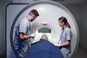
Breast MRI scans are a safe, accurate and non-invasive procedure to view breast tissue more completely. They’re just as important, though, for how they help spot abnormalities with unmatched precision. Here’s what you can expect, and what a vital role technologists play in the experience. It describes how we work to provide a safe and comfortable experience.
What Happens During the Scan
The procedure starts with patients positioned prone on a cushioned table specially made for breast imaging. The table has openings for the breasts to ensure stability and the best possible position. MRI technologists help patients through every step, describing what will happen and encouraging patients to ask any questions they may have.
Since the scan can take 30-45 minutes, patients need to be still to get clear images, which can be difficult for many patients. Doctors administer contrast agents through an IV to improve contrast between different types of tissue. This can be particularly helpful when focusing in on areas with compromised blood flow.
These agents significantly improve the sensitivity of malignant lesion detection. Malignant lesions typically have lower ADC values compared to benign lesions.
Comfort Features for Patients
Today’s state-of-the-art MRI machines put patients’ comfort and experience first. Open-bore systems provide additional space to reduce claustrophobic sensations, and adjustable padding improves comfort. Patients may choose to enjoy music throughout the scan, which helps foster a peaceful atmosphere.
Relaxation techniques, including deep or controlled breathing and guided imagery or visualization, may be used as well. These features combined help diminish anxiety, fostering a more pleasant experience.
Safety Measures and Noise Management
As with any other procedure, ensuring patient safety is of the utmost importance. MRI technologists are trained to closely monitor patients and to respond to any needs or issues quickly and effectively. You will be given earplugs or noise-canceling headphones to help with the machine’s loud noises.
It can be overwhelming and difficult to concentrate. Advanced noise-reduction technology helps create an even quieter environment. These precautions, together with continuous dialogue with technologists, make for a safe and comforting experience.
Interpreting Breast MRI Results
Breast MRI results are crucial in the early detection, diagnosis, and treatment planning of breast health. Radiologists, highly trained in the nuances of this science, carefully look through these images to find the essential information hidden within. Their interpretation determines the trajectory of patient care and helps to provide each patient with the individualized, accurate, and timely medical direction they need.
How Radiologists Analyze Images
Radiologists use a combination of techniques to interpret MRI images effectively. They evaluate signal intensity, enhancement patterns, and tissue morphology, often relying on T2-weighted imaging for additional clarity. Comparisons with prior imaging studies provide context, helping to distinguish changes over time.
Collaborative efforts between radiologists and oncologists further enhance accuracy, combining imaging insights with clinical expertise for comprehensive evaluations.
Understanding Normal Versus Abnormal Findings
Normal breast tissue should have a homogeneous structure with no abnormalities. Other abnormal features like persistent enhancement or irregular margins can suggest the presence of a malignant tumor. For instance, cancers less than 0.4 inches are often seen on MRI, highlighting its sensitivity.
Follow-ups, in the case of abnormalities, are necessary to inform subsequent management.
Next Steps After Receiving Results
Follow up actions may lead to breast biopsies or more advanced imaging. It is important for patients to come with questions prepared to discuss their results and what they mean. Quick, accurate communication helps patients make the most informed decisions possible and get on the best path to care.
Addressing Potential Risks and Concerns
Breast MRI, particularly with the advent of high-field sophisticated technology, provides the most sensitive imaging modality available to detect abnormalities. Yet, like any medical procedure, it is important to understand potential risks and the protocols in place to address them. This makes certain that patients are well informed, comfortable and confident in the care they are receiving.
Common Risks Associated with Breast MRI
One of the main potential risks has to do with gadolinium-based contrast agents. These agents improve image quality by accentuating variations in tissue. Though reactions are uncommon, some people may have a reaction that leads to mild nausea or a localized rash. Severe allergic reactions are very rare but call for readiness.
Patients with compromised kidney function need to be screened and risked, because the use of gadolinium has been shown to be harmful towards those with compromised renal function. Claustrophobia is a third risk associated with the MRI, considering the enclosed design of standard machines. This can create distress or apprehension for certain patients.
By screening for claustrophobia ahead of time, healthcare providers can recommend alternative solutions such as open MRI machines or offer calming techniques. Individual risk factors, such as existing health conditions or prior allergic reactions, further underscore the need for personalized assessment before scans. A thorough pre-scan evaluation minimizes risks and ensures the procedure is tailored to each patient’s needs.
Managing Claustrophobia During the Scan
For patients who experience anxiety in tight spaces, these approaches make a difference. Often relaxation techniques like breathing exercises or guided visualization work to instantly reduce anxiety. Today, many facilities have started to provide open MRI machines that minimize the feeling of confinement.
Sedation is the other alternative for the most severe cases, though it requires extra forethought. Clear communication and expectations are very important throughout the scan. Patients must not hesitate to speak up about their concerns with the MRI team.
The medical team will offer plenty of reassurance and remain in continuous communication via the intercom system throughout the procedure. Even basic interventions, such as giving patients headphones with relaxing music, can help create a more positive environment.
Addressing Concerns About Contrast Agents
Contrast agents hold a very central role in improving MRI images. Greater visibility allows for earlier and more accurate detection of abnormalities. For instance, MR imaging demonstrated the presence of recurrent invasive ductal carcinoma in a 73-year-old female. This level of precision frequently results in improved performance.
To prevent potential adverse events, healthcare providers observe patients for a period of time during and after the administration of contrast agents. Current protocols mandate screening for allergies and kidney function before administration.
We urge patients to report any past history of adverse reactions or current medical issues to their healthcare provider to help inform clinical decision-making.
Types of Breast MRIs
Breast MRIs provide state-of-the-art imaging technology to meet a wide range of imaging needs for breast health. These MRIs are designed for different goals, such as early detection, diagnostic clarity, or tracking treatment progress. Below is a categorized overview:
|
Type of Breast MRI |
Purpose |
|
|---|---|---|
|
Screening MRI |
Detect breast cancer in high-risk individuals |
Identifies abnormalities missed by mammograms, suitable for women under 40 or with dense tissue |
|
Diagnostic MRI |
Investigate specific symptoms or ambiguous mammogram findings |
Pinpoints size, location, and multiple lesions, evaluates lymph nodes for cancer cells |
|
Monitoring MRI |
Assess treatment response and guide ongoing cancer therapy |
Tracks tumor changes, ensures effective treatments, and detects new cancer in the opposite breast |
Screening MRI for High-Risk Individuals
Specialists know that screening MRIs are most valuable for those with higher-than-average risk of developing breast cancer. High-risk populations include women who have a significant family history of breast cancer. They are increasingly expanding to include carriers of BRCA1 or BRCA2 genetic mutations, and women with dense breast tissue.
This additional technique is extremely valuable for detecting small lesions. It’s capable of identifying cancer cells in underarm lymph nodes that a regular mammogram or ultrasound may overlook.
With access to the right data and insights, personalized screening protocols can be deployed to improve early detection and patient outcomes. For instance, abbreviated breast MRI — a newer, faster method — enables high-risk women to have more frequent screenings each year.
Diagnostic MRIs are highly effective for younger people. They offer a powerful preventative approach to identify and address issues before the typical mammogram starting age of 40.
Diagnostic MRI for Specific Symptoms
Diagnostic MRIs are used when addressing particular breast symptoms or inconclusive mammogram results. For instance, in cases where a mammogram cannot differentiate between scar tissue and a potential lesion, MRI provides detailed imaging clarity, even for patients with implants.
It evaluates the size, shape, and distribution of lesions, determining whether multiple areas within the breast are involved. It’s the targeted imaging capabilities of diagnostic MRIs that help ensure every patient undergoes the most precise and thorough evaluations.
Women newly diagnosed with breast cancer receive many more benefits than harms from MRIs. These scans are crucial for catching cancer in the contralateral breast, which occurs in approximately 10 percent of cases.
By rectifying such scenarios with precision, diagnostic MRIs facilitate well-informed treatment choices.
Monitoring Treatment Progress with MRI
Monitoring MRIs are essential to understanding how treatments for breast cancer are working. These scans give us very precise measurements of how tumors are responding to therapies such as chemotherapy and radiation.
In particular, they provide insight into changes in tumor size and tumor composition. These insights inform future treatment plans and keep interventions on track with what patients truly need.
Regular imaging is equally vital in detecting new abnormalities post-treatment, particularly in the opposite breast. The detailed imaging capabilities of breast MRIs allow physicians to monitor changes over time, ensuring that no recurrence or progression is overlooked.
Patients relying on ongoing therapy benefit from this continuous evaluation, improving their overall prognosis.
Future of Advanced High-Field MRI in Mammography
The integration of advanced high-field MRI into mammography practices is a promising leap ahead in breast cancer detection, prevention, and treatment. This approach capitalizes on advanced imaging technologies. It provides unmatched sensitivity and specificity, particularly for those with greater risk factors or dense breast tissue.
The potential applications extend beyond conventional screening. They create pathways for new breakthroughs in advanced, personalized care and broaden the field’s work in oncology.
Emerging Technologies Enhancing MRI Systems
These high-field MRI systems continue to advance with new breakthroughs that improve imaging quality and patient comfort. Such as the integration of diffusion-weighted imaging to better distinguish between malignant and benign tissues. This is particularly important when time limitations only allow for a few imaging sequences.
Recent studies using dynamic contrast-enhanced MRI (DCE-MRI) have increased sensitivity rates to a remarkable 91%. This is a dramatic improvement from the average 33% sensitivity of mammography. These innovations enhance diagnostic precision, lowering the risk of false positives and preventing unnecessary biopsies.
Ongoing research will be needed to fully optimize these tools. High-field MRI has increased the sensitivity by allowing the detection of tumors down to 0.03 inches (0.8 mm). This development is a huge asset in the fight against the most aggressive cancers.
Advancements such as noise reduction and faster scan times are dramatically changing the patient experience. With these new technologies come improvements to accessibility and comfort, opening up procedures to more individuals and improving their experiences.
This blend of novel accuracy and new improved usability through automation highlights the value of continued investment in MRI R&D.
Potential for Personalized Breast Care
The optimization of high-field MRI is a perfect demonstration of personalized medicine. By tailoring imaging protocols to individual risk factors, such as genetic predisposition or family history, clinicians can offer more precise and targeted care. Women with genetic predispositions, like BRCA1 carriers, reap tremendous benefits from these innovations.
Compared to standard mammography, high-field MRI provides a much more sensitive screening alternative. Women 40 years and younger typically have difficulty with traditional methods given the density of breast tissue. MRI offers a trusted solution for them.
By taking this personalized approach we’re helping to ensure that more women are detected early when treatment is most effective, leading to better long-term outcomes. By customizing imaging strategies, patients can be assured that they are receiving care that is appropriate to their individual needs.
This strategy eliminates unnecessary procedures and focuses resources on interventions that work. The incremental cost per quality-adjusted life year (QALY) for BRCA1 carriers was $14,005. This graphic illustrates the economic viability of incorporating high-field MRI into routine screening for high-risk groups.
Expanding Applications in Cancer Detection
High-field MRI has already been shown to be invaluable for early detection of breast cancer. Its advantages extend further than breast cancer, helping to identify other cancers as well. Emerging research emphasizes its utility in detecting cancers in difficult-to-screen regions, like the brain, liver, and prostate.
It is able to find smaller tumors, increasing the chances of an early diagnosis. This is essential for increasing survival in the majority of cancers. Its sensitivity to smaller lesions makes it an important tool. This advanced capability is key to monitoring high-risk patients and informing and planning targeted treatments.
Future directions for high-field MRI in oncology include integrating AI-driven analysis to further streamline image interpretation and improve diagnostic accuracy. Such innovations would increase its contribution to routine cancer care, ensuring that it remains an invaluable resource in every robust oncology practice.
Current studies are investigating its use as a non-invasive replacement for biopsies, sparing patients discomfort and procedural risk.
Conclusion
Advanced high-field MRI presents a non-ionizing, clear, detailed, and versatile alternative for breast imaging. It produces clearer pictures, allowing physicians to identify abnormalities sooner and more precisely. What makes this technology unique is its tremendous sensitivity to detect the earliest changes in breast tissue. The entire process, from preparation through results, is designed with patient comfort and care in mind. Though no test is without limitation, this approach to advanced high-field MRI for mammograms offers an innovative and trusted solution for breast health monitoring.
As the technology expands, it offers stronger outcomes and wider applications. Knowing these innovations puts you in control so you can feel confident you’re making the best health care decisions for yourself. Discuss with your doctor to determine if advanced high-field MRI is right for you. Your well-being deserves the most cutting-edge tools that exist.
Frequently Asked Questions
What is an advanced high-field MRI for mammograms?
An advanced high-field MRI, combined with computer-aided detection technology, uses high-powered magnetic fields to produce detailed, cross-sectional images of breast tissue. This tool drastically improves the detection of significant abnormalities compared to traditional imaging modalities. As a result, it is far more effective at screening for and diagnosing breast cancers.
What are the benefits of advanced high-field MRI for breast imaging?
This state-of-the-art technology allows for superior-quality, more accurate images. It can detect tiny anomalies that regular mammograms could overlook. This added capability improves early diagnosis, treatment and prevention of breast diseases.
Who should consider a breast MRI?
A breast MRI is recommended for individuals with a high risk of breast cancer, dense breast tissue, or inconclusive mammogram results. Your doctor will decide together with you if it’s the right choice for you.
How should I prepare for a breast MRI?
Do not wear anything metal, including jewelry or clothing with metal components. Follow all fasting instructions prescribed by your physician, and notify personnel of any implants, allergies or pregnancy.
Is a breast MRI painful?
No, in fact the procedure is quite comfortable. Lie perfectly still in the MRI scanner. While this process might seem intimidating, we promise you, this experience is not physically painful.
Are there any risks associated with advanced high-field MRI?
MRI is considered very safe. They are not ideal for patients with metal implants, pacemakers, or extreme claustrophobia. Be sure to always tell your doctor your full medical history in advance.
What is the future of advanced high-field MRI in mammography?
Looking to the future, imaging will be even faster, results even clearer, with AI-assisted diagnostics speeding up the process even more. These advances are meant to increase breast cancer detection rates and better personalize the treatment.