Discover the groundbreaking world of tomographic mammography, revolutionizing breast cancer detection. Traditional mammograms provide a two-dimensional view, while tomographic mammography, also known as breast tomosynthesis exams, offers a three-dimensional perspective, enhancing accuracy and early detection rates. By capturing multiple images from various angles, this advanced 3d mammography technology provides detailed insights, especially in dense breast tissue cases where traditional mammography methods may fall short. Stay ahead in your breast health journey with tomographic mammography’s precise imaging capabilities, offering a comprehensive approach to screening and diagnosis using mammogram images. Embrace the future of breast cancer detection with this cutting-edge 3d mammography tool that sets a new standard in women’s healthcare.
Key Takeaways
-
Breast Tomosynthesis Actionable Insight: Consider requesting tomographic mammography for a more detailed and accurate breast imaging, especially if you have dense breast tissue or a high risk of breast cancer.
-
Procedure Preparation Recommendations: Prior to the test, ensure to follow specific guidelines like avoiding deodorant, lotion, or perfume on the day of the scan to obtain clear and precise results.
-
Benefits Reminder: Breast tomosynthesis (3D mammography) offers advantages such as improved cancer detection rates and reduced call-backs for additional testing, enhancing the overall accuracy of breast cancer screening.
-
Risk Awareness: While tomographic mammography involves minimal additional radiation exposure compared to traditional mammograms, it’s crucial to discuss any concerns with your healthcare provider to make an informed decision.
-
Empowerment through Knowledge: Understanding the procedure and its benefits empowers individuals to actively participate in their breast health management, fostering early detection and potentially better treatment outcomes.
-
Personalized Care Emphasis: Tailoring breast imaging approaches like tomographic mammography based on individual needs, age, and risk factors can lead to more personalized and effective screening strategies.
Understanding Breast Tomosynthesis
What is Breast Tomosynthesis
Breast tomosynthesis is an advanced imaging technique that generates three-dimensional images of the breast, also known as a mammogram. It plays a crucial role in improving early detection and diagnosis of breast diseases by capturing multiple mammogram images from various angles, enhancing diagnostic accuracy.
Common Uses
-
Breast tomosynthesis is utilized for both screening and diagnostic purposes in breast health.
-
It effectively reduces false positives, especially in women with dense breast tissue.
-
Professional organizations recommend annual screening starting at age 40 to enhance early detection.
Screening vs Diagnostic Mammography
Screening mammography targets asymptomatic women, while diagnostic mammography focuses on symptomatic individuals. Breast tomosynthesis enhances both types by improving accuracy and enabling a more detailed examination. Tailored approaches based on age, individual risk factors, and symptoms are essential.
Equipment Overview
-
The digital mammography unit for breast tomosynthesis employs low-dose x-ray systems and computer reconstructions.
-
Breast compression is crucial for obtaining high-quality images that aid in accurate diagnosis.
Procedure Overview
How It Works
Breast tomosynthesis, also known as tomographic mammography, involves capturing multiple images of the breast from various angles. Computer algorithms then reconstruct these images into a 3D format for detailed examination. This method offers superior clarity and accuracy compared to traditional 2D mammography.
Performing the Test
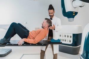
To conduct a breast tomosynthesis test, patients are positioned in front of the imaging machine. The equipment moves in an arch over the breast, capturing images at different angles. Patient cooperation is crucial during this process to ensure optimal image quality. Following safety protocols such as remaining still helps in obtaining precise and reliable results.
Experience During and After
During the imaging process, patients may experience slight pressure or compression on the breast, which is necessary for image quality. However, this discomfort is minimal and lasts only briefly. Patients should expect the procedure to last around 10-15 minutes per breast. After the test, patients receive guidance on any immediate follow-up steps, if required.
Interpreting Results
Radiologists analyze the 3D images produced by breast tomosynthesis to detect any abnormalities or signs of breast cancer. Results can vary from normal findings to indications that further tests are needed for conclusive diagnosis. It is vital for patients to discuss their results with their healthcare providers for appropriate guidance and next steps.
Preparation for the Test
How to Prepare
Patients should inform their doctor about any relevant medical history before the examination. This includes details about previous breast surgeries or implants. It is crucial for the medical team to have a comprehensive understanding of the patient’s health background to ensure a successful exam.
Avoid using certain products like deodorants on the day of the test. These products can interfere with the imaging process, affecting the quality of the results. Patients are advised to follow specific guidelines provided by the healthcare facility to prepare adequately for tomographic mammography.
Scheduling the exam when breasts are less likely to be tender is recommended. For many women, this typically means scheduling the test after menstruation. Breast tenderness can impact the comfort level during the exam, so choosing an optimal time can enhance the overall experience.
What to Expect on Test Day
On the day of the exam, patients should be prepared for the logistics of arriving at the facility. This includes check-in procedures, which may involve providing personal information and insurance details. Arriving early can help streamline this process and reduce any unnecessary stress before the examination.
Bringing prior mammogram images, if available, is essential. These images serve as valuable reference points for comparison with current results. Having these images on hand can aid radiologists in detecting any changes or abnormalities in breast tissue over time.
Patients should expect to encounter a professional and supportive environment during the imaging process. Medical staff will be present to assist and guide patients through each step of the examination. Their expertise ensures that patients receive high-quality care throughout the entire procedure.
During the Procedure
The technician will carefully position the patient to achieve optimal imaging results. This involves adjusting equipment and ensuring proper alignment for accurate scans. Patients should follow instructions from the technician to facilitate a smooth and efficient imaging process.
Breast compression is a standard part of tomographic mammography and is necessary for obtaining clear images. While it may cause slight discomfort, compression helps spread out breast tissue evenly, reducing overlap and enhancing image quality. Patients should communicate any concerns during this stage for adjustments as needed.
Patients can rest assured that tomographic mammography is a quick procedure that utilizes low radiation doses. The process is designed to be efficient and safe, minimizing exposure while capturing detailed images of breast tissue for analysis.
After the Procedure
After completing the examination, patients can expect results within a specified timeline determined by the healthcare facility. The waiting period may vary based on factors such as workload at the facility and result interpretation times.
In cases where additional imaging or tests are required based on initial findings, healthcare providers will discuss next steps with patients. It is important for patients to follow through with any recommended follow-up procedures to ensure a comprehensive evaluation of their breast health.
Patients are encouraged to ask questions or raise any concerns they may have following tomographic mammography. Clear communication with healthcare providers can address uncertainties and provide reassurance regarding the examination process and results.
Benefits and Risks
Benefits of Tomosynthesis
Tomographic mammography offers increased detection rates for small breast cancers through 3D imaging, enhancing the chances of early diagnosis and treatment. This technology allows for a more detailed examination, improving the chances of detecting abnormalities that may be missed in traditional 2D mammograms.
The utilization of tomosynthesis has shown a significant reduction in false positive results, leading to fewer unnecessary biopsies for patients. This decrease in false alarms helps alleviate anxiety and stress for individuals awaiting further tests or procedures, promoting a more efficient and accurate diagnostic process.
Improved ability to identify abnormalities in dense breast tissue is another crucial advantage of digital breast tomosynthesis (dbt). By providing clearer images and better visualization of dense areas, tomosynthesis enhances the accuracy of detecting potential issues, especially in women with denser breast tissue.
Risks Involved
While tomographic mammography is a valuable tool in breast cancer screening, there are certain risks associated with the procedure. Patients undergoing this imaging technique are exposed to a slight risk of radiation, although the benefits of early cancer detection typically outweigh this minimal risk.
One potential risk is the occurrence of false positive results, which can lead to unnecessary worry and stress for patients. The emotional impact of receiving a false positive result can be significant, highlighting the importance of clear communication between healthcare providers and individuals undergoing screening.
In some cases, follow-up tests may be required due to initial findings from tomosynthesis, adding an extra layer of uncertainty and potential anxiety for patients. It is essential for healthcare professionals to provide support and guidance throughout the entire screening process to ensure optimal care and peace of mind for individuals.
Minimizing Radiation Exposure
To mitigate the risks associated with radiation exposure during tomographic mammography, healthcare providers often recommend the use of low-dose x-ray technology. This approach helps minimize the amount of radiation absorbed by the body while still obtaining high-quality images for accurate diagnosis.
Adhering to guidelines for regular screenings is crucial in balancing benefits and risks associated with breast tomosynthesis. By following recommended screening intervals, individuals can ensure early detection of any abnormalities while minimizing unnecessary exposure to radiation over time.
Educating patients on the cumulative effects of radiation from repeated screenings is essential for informed decision-making regarding their healthcare. Understanding the long-term implications of radiation exposure empowers individuals to make educated choices about their screening frequency and overall health management.
Limitations
Despite its advantages, breast tomosynthesis also has limitations that need to be considered. Availability and cost factors may limit access to this advanced imaging technology for some individuals, impacting their ability to benefit from its enhanced diagnostic capabilities.
There is a potential for false negatives with breast tomosynthesis, particularly in certain breast types where abnormalities may not be as easily detectable. Healthcare providers must be aware of these limitations and consider additional screening methods when necessary to ensure comprehensive care for all patients.
Ongoing research is essential to improve technology and diagnostic capabilities related to breast tomosynthesis. By advancing the accuracy and effectiveness of this imaging technique, researchers can address current limitations and enhance its overall utility in detecting breast abnormalities at an early stage.
Closing Thoughts
Breast tomosynthesis, also known as 3D mammography, offers a more detailed view of breast tissue, enhancing the accuracy of detecting abnormalities. This advanced technology reduces the need for additional imaging and provides a comprehensive evaluation of your breast health. Understanding the benefits and risks associated with tomographic mammography empowers you to make informed decisions about your health.
Prioritizing regular breast screenings is crucial for early detection and improved treatment outcomes. Take charge of your well-being by scheduling a consultation with your healthcare provider to discuss the suitability of tomographic mammography for you. Your proactive approach to breast health can lead to timely interventions and better prognoses. Stay informed, stay vigilant, and prioritize your health above all else.
Frequently Asked Questions
Is tomographic mammography the same as traditional mammography?
Tomographic mammography, also known as breast tomosynthesis, is an advanced form of traditional mammography. While both methods involve imaging the breast tissue, tomosynthesis captures multiple images from different angles to create a 3D view, offering clearer results.
What is the importance of understanding breast tomosynthesis?
Understanding breast tomosynthesis helps patients comprehend the benefits of this advanced imaging technique over traditional mammography. It enables better detection of abnormalities in breast tissue and provides more accurate results, leading to early diagnosis and improved treatment outcomes.
How should one prepare for a tomographic mammography test?
Preparation for a tomographic mammography test typically involves wearing comfortable clothing and avoiding deodorants or lotions on the day of the exam. Patients may be asked to undress from the waist up and wear a gown during the procedure for optimal imaging results.
What are the benefits of undergoing tomographic mammography?
Tomographic mammography offers benefits such as improved detection of small lesions, reduced false positives, and enhanced visualization of breast tissue layers. This leads to increased accuracy in diagnosing breast cancer at an early stage, potentially improving treatment options and outcomes.
Are there any risks associated with tomographic mammography?
While tomographic mammography is considered safe, some risks may include exposure to low levels of radiation during the procedure. However, the benefits of early detection and accurate diagnosis of breast abnormalities generally outweigh the minimal risks associated with radiation exposure.