-
-
What are the key mammography views explained in the blog post?
-
How can positioning techniques help in addressing common challenges?
-
What benefits can healthcare professionals gain from enhancing their positioning skills?
-
Why is patient comfort important in mammography positioning?
-
What advanced tips are provided in the blog post to enhance mammography positioning?
-
Want to master the art of mammography positioning techniques for accurate results, including normal mammogram and craniocaudal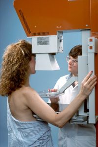 view? Dive into the world of mammography positioning techniques with us. Uncover the secrets to achieving optimal imaging outcomes and enhance your skills in this critical aspect of radiology practice with advanced mammography positioning course technique and mammography resources. Discover how to position patients effectively, ensuring high-quality mammograms every time. Stay tuned as we explore the nuances of positioning techniques that can make a significant difference in diagnostic accuracy and patient care. Ready to elevate your mammography game? Let’s delve into the essential strategies and best practices that will set you apart in the field.
Key Takeaways
-
Mastering mammography positioning techniques is crucial for accurate breast imaging.
-
Understanding the different mammography views and their significance is essential for quality imaging outcomes.
-
Tailoring positioning for special cases like implants or lymphedema ensures comprehensive patient care.
-
Overcoming common challenges in positioning, such as dealing with dense breast tissue, improves image quality.
-
Prioritizing patient comfort during mammography procedures leads to better overall experiences and compliance.
-
Continuously refining and incorporating advanced positioning tips elevates the quality of mammography exams.
Understanding Mammography
Importance of Positioning
Proper patient positioning is crucial for accurate mammography imaging. It plays a pivotal role in achieving clear visualization and high-quality interpretation. Adequate techniques significantly reduce motion artifacts and overlapping tissue, enhancing diagnostic accuracy.
Positioning is vital in mammography as it ensures that the breast tissue is spread out evenly, allowing for a comprehensive view. Incorrect positioning can lead to distorted images and potential missed abnormalities. Technologists must follow precise protocols to achieve optimal results.
The accuracy of mammogram results heavily relies on the technologist’s skill in positioning the patient correctly. Even minor errors in positioning can impact the quality of the images produced, affecting the radiologist’s ability to detect abnormalities early.
Key Views Overview
The mediolateral oblique (MLO) and craniocaudal (CC) views are essential in standard mammography screenings. The MLO view provides a side angle of the breast, while the CC view offers a top-to-bottom perspective. Together, they ensure a thorough examination of breast tissue.
These key views allow radiologists to visualize different areas of the breast, ensuring no abnormalities are overlooked. By capturing images from multiple angles, technologists can identify subtle changes in breast tissue density or structure that may indicate early signs of cancer.
Obtaining both MLO and CC views is critical for a comprehensive screening examination. Each view offers unique information about the breast tissue, enhancing the detection of abnormalities. The combination of these views increases the chances of detecting breast cancer at an early stage.
Mammography Views Explained
MLO View
A mediolateral oblique (MLO) view is crucial in mammography for capturing breast tissue from different angles. Proper positioning of the breast and compression paddle is essential for optimal MLO images. Assessing the adequacy of MLO acquisition involves evaluating the visibility of the pectoralis muscle and inframammary fold.
The MLO view is vital as it allows radiologists to visualize breast tissue near the chest wall, which might not be visible in other views. Positioning the breast correctly involves pulling it towards the chest wall while ensuring even compression with the paddle. Inadequate positioning can lead to overlapping tissue, affecting image clarity.
-
Pros:
-
Provides a comprehensive view of breast tissue.
-
Helps detect abnormalities closer to the chest wall.
-
-
Cons:
-
Requires precise positioning for accurate results.
-
CC View
The craniocaudal (CC) view plays a significant role in mammography by capturing a top-down image of the breast. It complements the MLO view by providing a different perspective for a thorough examination. Proper patient positioning is critical to obtaining clear and accurate CC images.
CC views are essential for detecting abnormalities in the upper portion of the breast that may not be visible in other views. When combined with the MLO view, radiologists can assess breast tissue comprehensively, improving detection rates. Ensuring proper patient positioning minimizes image distortion and enhances diagnostic accuracy.
-
The CC view offers a unique angle for examining breast tissue.
-
Combining CC and MLO views enhances diagnostic capabilities.
Positioning for Special Cases
Implant Displaced Views
Implant displaced views in mammography involve adjusting the positioning to visualize breast tissue effectively when implants are present. This technique is crucial for accurate breast cancer screening, especially in women with breast implants and normal mammogram. Challenges arise due to the interference of implants with breast tissue visibility during a normal mammogram.
Breasts with implants require specific considerations during imaging to ensure accurate results, including normal mammogram. The presence of implants can obscure breast tissue, making it challenging to detect abnormalities. Radiographers must adjust the technique to displace the implant adequately and visualize all breast tissue layers.
To achieve optimal imaging quality in cases of implant displacement, radiographers should use Eklund technique or implant displacement views. These techniques involve pushing the implant back against the chest wall to expose as much breast tissue as possible. It is essential to communicate effectively with patients and provide clear instructions during the process.
Challenging Patients Tips
Handling challenging patients during mammography positioning requires patience and effective communication skills. Empathy and understanding are key to managing anxious or difficult patients. Radiographers should explain each step of the procedure clearly to alleviate patient concerns.
Ensuring patient comfort and cooperation is vital for obtaining high-quality mammograms. Radiographers can offer blankets or cushions for comfort and adjust the positioning equipment as needed. For patients with specific needs, such as mobility issues, customized positioning techniques may be necessary.
Guidance on adapting positioning techniques for challenging patients includes allowing breaks between positioning adjustments and providing reassurance throughout the procedure. Maintaining a calm and supportive environment can help reduce patient anxiety and improve cooperation.
Positioning Techniques for Common Challenges
Large Breasts
Imaging large breasts in mammography requires special considerations to ensure accurate results. Proper compression is crucial for achieving clear images, especially in dense breast tissue. Technologists must use specific techniques to position the breasts adequately on the imaging plate.
To overcome challenges related to imaging large breasts, technologists may need to adjust the compression level based on breast size and density. Utilizing angled positioning techniques can help capture all breast tissue effectively. Ensuring that the breast is evenly compressed from top to bottom is vital for optimal image quality.
Effective imaging of large breasts relies heavily on proper compression and positioning. Inadequate compression can result in obscured or overlapping breast tissue, leading to inaccurate readings. Technologists must prioritize achieving uniform compression across the entire breast for thorough and precise imaging.
Small Breasts
When dealing with small breasts in mammography, technologists face unique challenges that require tailored solutions. Optimizing image quality in women with small breast sizes involves using specialized positioning techniques. The compression paddle must be adjusted to accommodate the smaller breast area adequately.
To enhance imaging outcomes for women with small breasts, technologists should focus on maximizing breast tissue visibility while maintaining patient comfort. Employing spot compression techniques can help target specific areas of interest within the breast for detailed examination. Tailoring positioning methods to suit individual breast sizes is essential for accurate results.
Accurate imaging of small breasts hinges on utilizing precise positioning techniques that allow for comprehensive coverage of the breast tissue. Technologists must pay close attention to detail when positioning smaller breasts to ensure that no areas are missed during the imaging process.
Limited Mobility
Accommodating patients with limited mobility during mammography positioning requires a compassionate and adaptable approach. Technologists should communicate effectively with patients to understand their mobility constraints and concerns. Alternative positioning approaches, such as seated or upright imaging, can be utilized for patients unable to stand comfortably.
Incorporating strategies that prioritize patient comfort and safety is paramount when adapting positioning techniques for individuals with limited mobility. Technologists must exercise patience and empathy while assisting patients with positioning adjustments to ensure a positive and stress-free experience during the mammography procedure.
Ensuring that patients with limited mobility feel supported and cared for throughout the imaging process is essential for maintaining their trust and cooperation. By implementing flexible positioning strategies and prioritizing patient well-being, technologists can facilitate a smooth and successful mammography session.
Enhancing Positioning Skills
ACR Guidelines Overview
The American College of Radiology (ACR) provides comprehensive guidelines for mammography positioning to ensure accurate and effective imaging. These guidelines serve as a crucial reference point for technologists involved in performing mammograms. The ACR guidelines encompass detailed instructions on patient positioning, compression techniques, and image quality standards.
Adhering to the ACR guidelines is essential for maintaining consistency and quality in mammography imaging. These guidelines outline specific recommendations for achieving optimal breast positioning, which is fundamental for accurate detection of abnormalities. By following the ACR standards, technologists can minimize errors and ensure that every mammogram meets the required quality benchmarks.
Key recommendations in the ACR guidelines include precise positioning of the breast within the compression paddle, appropriate angling for optimal tissue visualization, and adequate compression to enhance image clarity. Furthermore, the standards emphasize the importance of regular calibration of equipment and adherence to radiation safety protocols during mammography procedures.
EQUIP in Positioning
Enhancing Quality Using the Inspection Program (EQUIP) plays a vital role in improving mammography positioning practices. This program focuses on continuous quality improvement through ongoing assessment and feedback mechanisms. Technologists are key players in implementing EQUIP strategies to enhance imaging outcomes and ensure consistent quality in mammography.
EQUIP encourages technologists to actively engage in self-assessment and peer review processes to identify areas for improvement in positioning techniques. By participating in regular training sessions and performance evaluations, technologists can refine their skills and stay updated on the latest advancements in mammography positioning.
The role of technologists in EQUIP implementation is pivotal for enhancing overall imaging quality. Technologists are responsible for applying best practices in breast positioning, ensuring patient comfort during examinations, and maintaining compliance with established protocols. Through collaboration with radiologists and other healthcare professionals, technologists contribute significantly to achieving accurate and reliable mammography results.
Addressing Patient Comfort
Wheelchair Positioning
Wheelchair positioning for mammography presents unique challenges, especially in ensuring patient comfort and image quality. Adapting standard positioning techniques becomes crucial when dealing with patients in wheelchairs. To address this, specialized equipment such as adjustable height chairs and dedicated wheelchair platforms are utilized. These tools aid in achieving optimal positioning for accurate imaging results. Ensuring accessibility is paramount to accommodate patients with mobility limitations effectively.
-
Use adjustable height chairs and dedicated wheelchair platforms
-
Ensure accessibility for patients with mobility limitations
Prominent Abdomens
Imaging patients with prominent abdomens requires specific considerations to overcome challenges related to abdominal tissue interference. Techniques such as adjusting compression paddle pressure and angling the tube can help mitigate these challenges. Optimizing imaging quality in these cases involves fine-tuning equipment settings and employing patient-specific approaches. By customizing techniques to suit individual anatomical variations, healthcare providers can enhance diagnostic accuracy while prioritizing patient comfort.
-
Adjust compression paddle pressure and angling the tube
-
Customize techniques for individual anatomical variations
Advanced Positioning Tips
Screening Initiation Time
Initiating screening mammograms in asymptomatic women typically begins at age 40. Regular screenings every 1-2 years are recommended for early detection. Early screening is crucial for reducing breast cancer mortality rates.
Screening at the age of 40 allows for the detection of potential abnormalities before symptoms arise. Regular mammograms help in identifying cancerous growths at an early, treatable stage. Early detection significantly improves treatment outcomes and survival rates.
-
Pros:
-
Early detection of abnormalities
-
Improved treatment outcomes
-
-
Cons:
-
Potential false-positive results
-
Increased anxiety for patients
-
Additional Views Explained
Additional views in mammography serve to provide a more comprehensive assessment of breast tissue. These views are essential in cases where standard images may not capture all areas adequately. Supplementary views play a critical role in ensuring accurate diagnosis.
In certain scenarios, such as when there is overlapping tissue or unclear findings, additional views become necessary. These views offer different angles of the breast, aiding radiologists in making informed diagnostic decisions. Supplementary imaging helps in reducing the chances of missed or misinterpreted findings.
-
The rationale behind additional views:
-
Comprehensive assessment of breast tissue
-
Enhanced diagnostic accuracy
-
-
Specific scenarios requiring additional views:
-
Dense breast tissue
-
Suspicious findings on initial images
-
-
Role of supplementary views:
-
Providing a detailed evaluation of breast structures
-
Assisting in accurate diagnosis and treatment planning
-
Summary
You have now gained valuable insights into mammography positioning techniques, from understanding the basics to mastering advanced tips. By delving into various views, special cases, common challenges, and patient comfort considerations, you are better equipped to enhance your positioning skills. Remember, attention to detail and continuous practice are key in achieving optimal imaging results.
Take the knowledge you’ve acquired here and apply it in your practice. Strive for precision in positioning to ensure accurate diagnoses and improved patient outcomes. Stay updated on advancements in mammography techniques, and never underestimate the impact of your expertise on the quality of care provided. Keep refining your skills, and you will make a significant difference in the field of mammography.
Frequently Asked Questions
What are the key mammographic views and mammographic positioning explained in the blog post?
The blog post explains essential mammography views like craniocaudal (CC) and mediolateral oblique (MLO) views, crucial for breast imaging accuracy.
How can positioning techniques help in addressing common challenges?
Positioning techniques play a vital role in overcoming challenges like overlapping tissues, ensuring optimal breast compression, and achieving clear imaging results.
What benefits can healthcare professionals gain from enhancing their positioning skills?
Enhancing positioning skills leads to improved accuracy in detecting abnormalities, reducing the need for re-imaging, and providing better patient care through precise diagnosis.
Why is patient comfort important in mammography positioning?
Ensuring patient comfort during mammography positioning helps reduce anxiety, improves cooperation for accurate imaging, and enhances the overall experience, leading to higher patient satisfaction.
What advanced tips are provided in the blog post to enhance mammography positioning, including normal mammogram, technique, nipple, and oblique view?
The blog post offers advanced tips such as utilizing cone-shaped compression paddles, adjusting for breast implants, and employing specialized techniques for challenging cases to elevate mammography positioning proficiency.