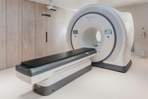
Did you know that advanced high-field MRI technology can capture images with a resolution up to 100 times higher than traditional MRI machines? This cutting-edge innovation is revolutionizing the field of medical imaging, providing unparalleled insights into the human body’s intricacies. With its high spatial resolution and sensitivity, advanced high-field MRI, powered by a strong magnet, is paving the way for more accurate diagnoses and personalized treatment plans. By harnessing the power of magnetic fields, MRI scanners offer a non-invasive yet incredibly detailed look inside the body, enhancing healthcare outcomes like never before.
High-Field MRI Basics
Understanding Technology

High-field MRI machines operate at higher magnetic field strengths, typically 1.5 Tesla or above. This advancement allows for enhanced image resolution and clarity, aiding in the detection of minute anatomical details. The technology behind these machines involves powerful magnets that align hydrogen atoms within the body, generating signals used to create detailed images.
The utilization of high-field MRI machines has revolutionized diagnostic capabilities by providing radiologists with sharper images for more accurate diagnoses. These machines offer improved contrast and spatial resolution, enabling healthcare professionals to detect abnormalities earlier and with greater precision in human studies. Understanding this advanced technology is crucial for healthcare providers to leverage the full potential of high-field MRI in enhancing patient care outcomes.
How MRIs Work
In high-field MRI machines, powerful magnets create a strong magnetic field that aligns hydrogen protons in the body along the magnetic field lines. When radio waves are applied, these protons absorb and release energy, producing signals that are detected by the machine’s receiver coils. This data is then processed to generate highly detailed cross-sectional images of the body’s internal structures.
MRI examinations are non-invasive procedures that do not involve ionizing radiation or magnetic field, making them safe for patients of all ages. The ability of MRI machines to produce detailed images without harmful radiation exposure makes them invaluable in diagnosing various musculoskeletal conditions such as ligament tears, joint abnormalities, and spinal cord injuries accurately.
Machine Components
High-field MRI machines consist of essential components like superconducting magnets, gradient coils, radiofrequency coils, and a computer system for image reconstruction. These superconducting magnets produce the main magnetic field required for imaging by circulating electrical currents through coils cooled to extremely low temperatures.
Compared to standard MRI machines, high-field systems have stronger gradient coils that enable faster imaging sequences and improved image quality. Radiofrequency coils transmit radio waves into the body to stimulate proton alignment and receive signals emitted during relaxation processes. Each component plays a vital role in capturing precise anatomical details and producing high-quality images essential for accurate diagnoses.
Benefits of High-Field MRI
Enhanced Imaging Quality
High-field MRI machines utilize stronger magnetic fields, resulting in clearer and more detailed images. These machines offer higher resolution, enabling healthcare providers to detect smaller abnormalities that may be missed with lower-field MRI scanners. The enhanced imaging quality plays a crucial role in improving the accuracy of diagnoses and treatment plans.
The impact of enhanced imaging quality is significant in various medical specialties, including neurology and oncology. For example, in neurology, high-field MRI can provide precise visualization of brain structures, aiding in the diagnosis of conditions such as tumors or multiple sclerosis. In oncology, the superior image quality allows for better delineation of tumors and surrounding tissues, guiding surgeons during procedures.
Key features contributing to improved imaging quality include higher signal-to-noise ratio (SNR), which enhances image contrast and clarity. High-field MRI machines offer advanced software algorithms that help reduce artifacts and improve overall image quality by minimizing distortions.
Faster Scan Times
High-field MRI machines are known for their efficiency in completing scans quickly, reducing overall scan times. This benefit is particularly advantageous for patients as it minimizes the time spent inside the machine, leading to a more comfortable experience. The faster scan times not only enhance patient comfort but also increase the throughput of imaging facilities.
Quick scans are essential in emergency situations where prompt diagnosis is critical for patient outcomes. By reducing scan durations, high-field MRI machines enable healthcare providers to obtain necessary information rapidly, facilitating timely interventions. Moreover, faster scan times contribute to improved workflow efficiency, allowing medical professionals to accommodate more patients within a given timeframe.
Multinuclear Imaging
Multinuclear imaging refers to the use of multiple nuclei, such as hydrogen and phosphorus, for acquiring images in high-field MRI machines. This technique offers unique insights into tissue characteristics and metabolic processes that cannot be obtained through conventional imaging methods. In specific diagnostic scenarios like cardiac imaging or spectroscopy studies, multinuclear imaging provides valuable information for accurate assessments.
The advantages of multinuclear imaging extend to research applications as well, allowing scientists to study cellular metabolism and molecular pathways with high precision. By leveraging different nuclei properties, healthcare providers can gain a comprehensive understanding of tissue composition and function. Multinuclear imaging plays a crucial role in advancing diagnostic capabilities and enhancing patient care through personalized treatment approaches.
High-Field MRI Applications
Neuroimaging Advances
Ultra-High-Field Applications
High-field MRI machines have revolutionized neuroimaging, but ultra-high-field MRI takes it a step further. These cutting-edge machines operate at field strengths above 3 Tesla, offering unparalleled image resolution and clarity. The unique benefits of ultra-high-field MRI include enhanced visualization of small structures in the brain, leading to more accurate diagnoses.
In advanced medical settings, ultra-high-field MRI is particularly effective in studying neurological disorders like multiple sclerosis and Alzheimer’s disease. These machines provide detailed images that aid in early detection and monitoring of disease progression. Ultra-high-field MRI enables researchers to explore brain connectivity and function with unprecedented detail.
Clinical Use in Neuroimaging
High-field MRI plays a critical role in neuroimaging, allowing healthcare providers to delve deep into the complexities of the brain. By utilizing high magnetic fields, these machines enhance the visualization of brain structures and abnormalities. This advancement significantly contributes to the diagnosis and treatment of various neurological conditions.
In clinical neuroimaging, specific imaging techniques such as functional MRI (fMRI) and diffusion tensor imaging (DTI) are commonly employed. These techniques provide valuable insights into brain activity, connectivity, and white matter integrity. High-field MRI’s ability to capture intricate details aids in understanding brain function and pathology.
Orthopedic Imaging
Ankle Sprain Example
Imagine a scenario where an athlete sustains an ankle injury during a game. Utilizing high-field MRI, healthcare professionals can accurately diagnose the extent of the injury through detailed imaging. By visualizing ligament tears or bone fractures with precision, early intervention can be initiated promptly.
High-field MRI’s capability to capture high-resolution images plays a crucial role in assessing ankle sprains effectively. Early diagnosis facilitated by this technology allows for timely treatment interventions, preventing long-term complications. In orthopedic practice, high-field MRI has become indispensable for accurate diagnosis and optimal management of musculoskeletal injuries.
Technical Challenges
B0 Inhomogeneity
B0 inhomogeneity refers to variations in the magnetic field strength within the imaging area during high-field MRI scans. These fluctuations can distort images and compromise diagnostic accuracy. Strategies such as shim techniques and gradient-echo sequences are employed to mitigate B0 inhomogeneity effects.
RF Power Deposition
RF power deposition, the energy absorbed by the patient’s body during high-field MRI scans, can have adverse effects on tissues. Safety precautions are crucial to prevent patient harm due to excessive RF power deposition. Optimizing scan parameters and limiting scan duration help minimize risks.
Relaxation Behavior Changes
In high-field MRI, relaxation behavior changes occur due to the stronger magnetic fields influencing tissue properties. These alterations impact image contrast and signal intensity, affecting the interpretation of MRI findings. Understanding relaxation behavior changes is essential for accurate diagnosis and treatment planning.
Overcoming Obstacles
Specialized RF Pulses
Specialized RF pulses in high-field MRI play a vital role in enhancing image quality and diagnostic accuracy. By utilizing these tailored radiofrequency pulses, radiologists can improve image contrast and clarity significantly. These pulses are specifically designed to target different tissues or structures within the body, allowing for enhanced visualization of abnormalities that may be challenging to detect with conventional MRI techniques.
Moreover, specialized RF pulses find critical applications in specific diagnostic scenarios such as neuroimaging and musculoskeletal imaging. In neuroimaging, these pulses help in highlighting subtle brain abnormalities, while in musculoskeletal imaging, they aid in detecting intricate joint pathologies. The ability to customize RF pulses based on the area of interest makes them a valuable asset in modern high-field MRI technology.
Chemical Shift Error
Chemical shift error is a common phenomenon encountered in high-field MRI that can lead to misinterpretation of images. This error occurs due to variations in the resonant frequencies of protons within different tissues. As a result, structures may appear displaced or distorted, posing challenges for accurate diagnosis.
To address chemical shift errors, radiologists employ various strategies such as adjusting imaging parameters, utilizing specialized sequences like fat suppression techniques, and implementing post-processing algorithms. By mitigating these errors effectively, healthcare providers can ensure reliable interpretation of MRI scans and provide patients with accurate diagnoses and treatment plans.
B1 Inhomogeneities
B1 inhomogeneities are another obstacle faced during high-field MRI scans that can impact image quality and diagnostic precision. These inconsistencies in the magnetic field’s strength across the imaging volume can lead to signal variations and distortions in the final images. As a result, anatomical structures may appear blurred or misrepresented.
Radiologists utilize advanced techniques like parallel transmission methods and multi-channel RF coils to address B1 inhomogeneities effectively. By optimizing RF transmission pathways and employing sophisticated hardware solutions, medical professionals can minimize these discrepancies and enhance the overall quality of high-field MRI images for improved patient care.
Safety and Regulatory Considerations
FDA Approval Process
Obtaining FDA approval for high-field MRI machines involves a rigorous regulatory process. Manufacturers must submit detailed data on the machine’s safety, performance, and efficacy. The FDA evaluates this information to ensure compliance with strict standards. The approval process typically includes preclinical testing, clinical trials, and post-market surveillance to monitor long-term safety.
Meeting the FDA requirements is crucial for ensuring the quality and safety of high-field MRI technology. The FDA sets specific standards for image quality, patient safety, and device performance. Compliance with these standards is essential to guarantee accurate diagnostic imaging and minimize potential risks to patients. FDA approval signifies that the MRI machine meets all necessary criteria for safe and effective use in clinical settings.
FDA approval plays a vital role in safeguarding patients undergoing high-field MRI scans. It ensures that the equipment meets stringent safety and performance guidelines. FDA-approved machines are more likely to provide reliable imaging results while minimizing potential hazards to patients.
Managing Physiologic Effects
Managing physiologic effects during high-field MRI scans is crucial for obtaining accurate diagnostic images. Patient motion, breathing patterns, and heart rate can introduce artifacts that affect image quality. To mitigate these effects, healthcare providers may implement techniques such as breath-holding instructions, patient coaching, or physiological monitoring during scans.
Physiologic effects have a direct impact on the quality of MRI images and the accuracy of diagnoses. Any movement or physiological changes can distort images and compromise diagnostic interpretation. Proper patient preparation, including education on scan procedures and expectations, can help reduce physiologic artifacts and improve overall imaging quality.
Effective patient preparation is essential to minimize physiologic artifacts during high-field MRI scans. Clear communication with patients about scan protocols, positioning requirements, and breathing instructions can enhance image clarity and diagnostic accuracy.
Implantable Devices Handling
Handling implantable devices in the context of high-field MRI poses unique challenges due to safety concerns. Patients with pacemakers, defibrillators, or other implants face potential risks during MRI scans. The strong magnetic fields generated by high-field MRIs can interact with metal implants, leading to device malfunction or tissue heating.
The risks associated with imaging patients with implantable devices highlight the need for specialized protocols and precautions. Healthcare providers must follow strict guidelines to ensure safe imaging practices for these individuals. This includes verifying device compatibility with MRI systems, monitoring patients closely during scans, and having emergency protocols in place in case of adverse events.
Protocols for safe imaging of patients with implantable devices involve thorough screening processes, device programming checks, and continuous patient monitoring throughout the scan. These measures are essential for preventing complications and ensuring patient safety during high-field MRI examinations.
Cost and Siting Considerations
Evaluating MRI Costs
High-field MRI scans’ costs are influenced by factors such as machine strength, image quality, and facility location. Evaluating MRI costs is crucial for patients to understand expenses and for healthcare providers to manage budgets effectively. Optimizing MRI costs involves strategies like bulk purchasing, maintenance planning, and energy efficiency improvements.
Siting Requirements
Setting up high-field MRI machines requires specific considerations including adequate space for the equipment, shielding against electromagnetic interference, and compliance with safety regulations. Meeting siting requirements ensures optimal MRI performance by minimizing interference, ensuring patient safety, and maintaining image quality.
Future of High-Field MRI
Technological Innovations
Recent advancements in high-field MRI technology have revolutionized medical imaging. These innovations have significantly enhanced imaging quality by providing clearer and more detailed images. Improved efficiency in scanning processes has reduced scan times, leading to quicker diagnoses and treatment plans for patients. The introduction of advanced software and hardware components has further optimized the patient experience during MRI procedures.
The future of high-field MRI is promising, with ongoing research focusing on enhancing image resolution, reducing scan times even further, and increasing patient comfort. Emerging technologies like artificial intelligence (AI) are being integrated into high-field MRI systems to automate image analysis and interpretation. This not only streamlines the diagnostic process but also ensures accuracy and consistency in results. Developments in contrast agents and specialized coils are expected to further improve image quality and diagnostic capabilities.
Regulatory Evolution
The regulatory landscape surrounding high-field MRI technology has evolved over the years to ensure patient safety and quality standards. Changes in regulations impact the development, manufacturing, installation, and operation of MRI machines. Compliance with these regulations is crucial for healthcare facilities to maintain accreditation and provide safe imaging services to patients.
Staying updated with regulatory evolutions is essential for healthcare providers and manufacturers involved in high-field MRI technology. Adhering to regulatory requirements ensures that equipment meets safety standards, performance criteria, and quality controls. Moreover, compliance with regulations helps in mitigating risks associated with equipment malfunctions or operational errors, safeguarding both patients and healthcare professionals.
Final Remarks
You’ve delved into the world of advanced high-field MRI, uncovering its basics, benefits, applications, technical challenges, and future prospects. Despite the hurdles faced in terms of safety, regulations, cost, and siting considerations, the potential for high-field MRI to revolutionize medical imaging is undeniable. Embracing this technology could lead to more accurate diagnoses, better treatment outcomes, and enhanced patient care.
As you ponder the insights gained from this exploration, consider how advancements in high-field MRI could shape the future of healthcare. Stay informed about the latest developments in this field and advocate for access to cutting-edge technologies that can improve medical practices. Your engagement and support can contribute to a brighter and healthier tomorrow.
Frequently Asked Questions
What are the advantages of high-field MRI technology?
High-field MRI offers superior image quality, faster scan times, and enhanced resolution compared to lower field strengths. This allows for more detailed insights into anatomical structures and improved diagnostic accuracy.
How is safety ensured in high-field MRI scans?
Safety in high-field MRI is maintained through strict adherence to protocols, screening procedures to identify contraindications, and continuous monitoring during scans to ensure patient comfort and well-being.
What are the key applications of advanced high-field MRI in clinical imaging, human scanners, and clinical settings?
Advanced high-field MRI is used in various medical fields such as neuroimaging, musculoskeletal imaging, cardiac imaging, and oncology. It enables detailed visualization of tissues and organs for accurate diagnosis and treatment planning.
How do technical challenges impact the use of high-field MRI systems in clinical imaging?
Technical challenges in high-field MRI include susceptibility artifacts, increased radiofrequency power deposition, and limitations in image uniformity. These challenges necessitate ongoing research and development efforts to enhance system performance.
What does the future hold for high-field MRI technology?
The future of high-field MRI technology involves advancements in gradient systems, RF coil design, and image reconstruction techniques. These innovations aim to further improve imaging quality, increase patient comfort, and expand the range of clinical applications.


